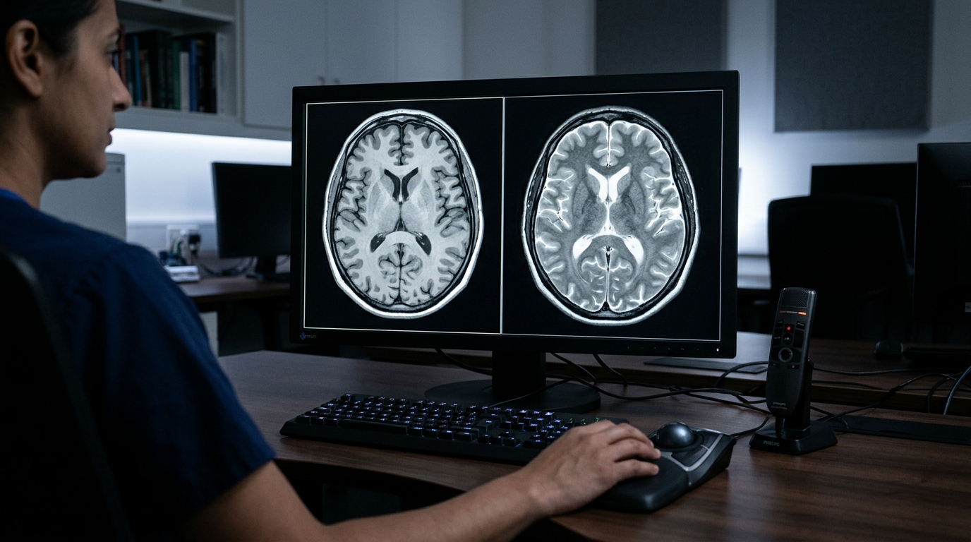
Magnetic resonance imaging serves as an indispensable, non-invasive modality within clinical diagnostics and medical research because of its exceptional soft-tissue contrast and capacity to delineate intricate anatomical structures. By exploiting the intrinsic magnetic properties of hydrogen atoms across human tissues, clinical magnetic resonance imaging relies on distinct radiofrequency pulse sequences to emphasize specific pathological and physiological characteristics. Standard examinations frequently acquire multiple complementary image sequences, including T1-weighted scans where fat appears hyperintense and cerebrospinal fluid appears dark due to long longitudinal relaxation times, and T2-weighted scans where water-rich tissues, inflammation, and edema appear bright because of extended transverse relaxation. Specialized sequences further broaden diagnostic capabilities: fluid-attenuated inversion recovery suppresses free fluid signals to reveal periventricular multiple sclerosis lesions, diffusion-weighted imaging captures microscopic Brownian motion of water to identify acute ischemic stroke, and T2*-weighted gradient-echo imaging detects field inhomogeneities from hemosiderin deposits and microbleeds. However, acquiring complete multi-sequence protocols directly from patients imposes significant burdens, including prolonged scanner durations, heightened operational expenses, substantial patient discomfort, and vulnerabilities to motion artifacts. In urgent clinical scenarios or resource-constrained settings, critical sequences are often omitted, leaving diagnosticians with incomplete information.
To overcome these clinical bottlenecks without subjecting patients to prolonged physical examinations or repetitive scanning sessions, medical imaging researchers have turned to deep learning algorithms capable of executing cross-contrast magnetic resonance image translation. In a comprehensive systematic review published in Discover Imaging, investigators Richard Acs and Hanqi Zhuang evaluated thirty carefully selected peer-reviewed studies published over a ten-year window up to May 19, 2024, assessing the architectural design, dataset composition, analytical metrics, and clinical utility of computational contrast synthesis. Across the surveyed literature, computational contrast translation encompasses three primary operational paradigms: unimodal one-to-one conversion from a single existing contrast to a target sequence, multimodal-to-unimodal synthesis combining complementary sequences such as T1-weighted, T2-weighted, and diffusion-weighted data into a single output like fluid-attenuated inversion recovery, and one-to-many translation where an algorithm reconstructs every missing contrast from a single available scan. Furthermore, specialized investigations have explored synthesizing artificial contrast-enhanced T1-weighted sequences directly from non-contrast acquisitions, an intervention that could potentially minimize or eliminate the clinical necessity of administering intravenous gadolinium-based contrast agents to vulnerable patient populations.
The methodological framework governing the systematic review entailed a rigorous multi-stage identification and screening workflow across major electronic scientific repositories. Richard Acs and Hanqi Zhuang searched IEEE Xplore, PubMed, and the Directory of Open Access Journals using predefined combinations of keywords including deep learning, MRI, synthesis, and generation, retrieving an initial corpus of 971 records, comprising 424 articles from IEEE Xplore, 484 articles from PubMed, and 63 articles from the Directory of Open Access Journals. The authors employed automated screening to filter records lacking magnetic resonance imaging designations in their titles, followed by independent manual screening by both reviewers against explicit exclusion criteria. Eligible investigations were required to apply deep learning to two-dimensional cross-contrast magnetic resonance synthesis; consequently, non-English publications, prior review papers, studies incorporating non-magnetic modalities such as positron emission tomography or computed tomography, investigations relying on raw k-space inputs or non-finalized quantitative parameter maps, and implementations operating on three-dimensional volume inputs were eliminated. Discrepancies were resolved through mutual consensus, yielding twenty-nine studies from database searches, with one influential investigation manually added from reference tracking, culminating in thirty analyzed publications.
Algorithmic architectures identified across the thirty reviewed studies follow distinct mathematical paradigms, beginning with foundational convolutional networks and autoencoders that establish direct non-linear mappings between paired source and target contrasts. Fully convolutional neural networks execute dense, pixel-wise predictions across image planes, while autoencoders leverage encoder-decoder pipelines to compress source contrasts into compact latent representations before decoding them into reconstructed target contrasts through reconstruction loss minimization. Serving as an architectural cornerstone across the discipline, the U-Net design features a symmetrical contracting downsampling path and an expansive upsampling path connected by cross-network skip connections. These skip connections transmit high-resolution localized spatial features directly from early encoder layers across to corresponding decoder stages, circumventing information loss and enabling precise anatomical preservation before final regression layers output the synthesized contrast. Beyond standalone deployment in resource-constrained environments, U-Nets serve extensively as generative sub-networks inside adversarial models and function as the core noise-predicting neural backbone within reverse diffusion architectures.
Generative adversarial networks constitute the most widely adopted framework in the cross-contrast synthesis literature, driven by their ability to generate sharp, visually realistic tissue textures through min-max game optimization between two opposing subnetworks. Within this adversarial paradigm, a generator network synthesizes artificial contrast samples from source inputs, while an adjacent discriminator network evaluates whether presented images originate from real clinical training distributions or synthetic algorithmic outputs, optimizing an objective function based on expected log-likelihoods. Because medical contrast translation requires strict anatomical fidelity rather than unconditional random image generation, researchers frequently employ conditional adversarial frameworks such as pix2pix, where source images constrain the generator loss. To bypass the severe clinical constraint of requiring perfectly aligned, identical-geometry image pairs, researchers widely deploy CycleGAN architectures, which incorporate two generators and two discriminators operating under cycle-consistency constraints to translate unpaired cross-contrast cohorts. Scaled variants like StarGAN further expand flexibility by mapping multiple contrast domains simultaneously using a single unified network, although adversarial models remain notoriously susceptible to training instability and mode collapse.
More recently, denoising diffusion probabilistic models, initially established in generative literature in 2020, have demonstrated state-of-the-art synthesis capabilities that systematically rival and occasionally exceed generative adversarial networks. Operating through a two-phase thermodynamic formulation, diffusion models define a forward process that incrementally degrades structural information by adding Gaussian noise over discrete time steps, and a parameterized reverse process that reconstructs clean data distributions from isotropic noise. In cross-contrast tasks, diffusion networks incorporate conditional guidance by concatenating source contrast slices directly to noisy inputs or injecting encoded multimodal latent vectors and explicit anatomical masks into the reverse sampling trajectory. Unlike adversarial architectures, diffusion models avoid discriminator instabilities and resist mode collapse, demonstrating remarkable fidelity in retaining microstructural tissue boundaries and delicate anatomical details. However, their reliance on extensive sequential reverse sampling steps imposes substantial computational latency, presenting practical hurdles for time-critical emergency applications unless accelerated through emerging fast-sampling algorithmic approximations.
Transformer-based architectures represent another rapidly advancing class of models reviewed in the literature, overcoming the fundamental constraint of convolutional receptive fields through multi-head self-attention mechanisms. While traditional convolutional filters operate locally, vision transformers divide two-dimensional magnetic resonance slices into sequences of distinct spatial patches, project them linearly into high-dimensional embeddings with added positional encodings, and calculate global attention weights across all anatomical regions simultaneously. Recent implementations highlight the architectural versatility of transformers in medical imaging: hybrid convolutional-transformer frameworks preserve local edge detection while modeling long-range spatial correlations, adjacent-slice contextual priors improve two-dimensional slice synthesis between T1-weighted and T2-weighted scans, and Edge-Preserving Masked Autoencoder pre-training reinforces structural boundaries necessary for surgical margins. Furthermore, multi-scale transformer frameworks have proven capable of handling arbitrary subsets of available contrasts to synthesize missing modalities dynamically, although heavy memory consumption and substantial training dataset requirements currently limit their widespread clinical deployment.
The empirical evaluation of synthetic contrast fidelity relies on standardized quantitative metrics, prominently featuring Peak Signal-to-Noise Ratio, Normalized Mean Square Error, and the Structural Similarity Index Measure. Peak Signal-to-Noise Ratio calculates logarithmic ratios between maximum possible pixel intensity values and the mean squared error separating synthesized signals from ground-truth scans, quantifying overall signal reconstruction accuracy. Normalized Mean Square Error scales the squared Euclidean norm of reconstruction errors by the ground-truth image norm to provide an invariant metric of total distortion. The Structural Similarity Index Measure evaluates perceptual congruence by modeling luminance, contrast, and structural covariance using stabilizing constants to prevent computational divergence in uniform tissue zones. In comparative benchmark evaluations conducted on the standardized BraTS 2018 dataset, U-Net models and generative adversarial networks exhibited comparable quantitative performance, routinely falling within a single standard deviation of each other across Structural Similarity and Peak Signal-to-Noise scores. Conditional diffusion models evaluated on identical benchmarks achieved among the highest recorded metrics, underscoring their capacity for superior structural restoration.
Despite strong numerical metrics, the review highlights critical discrepancies between mathematical similarity scores and real-world clinical interpretability, underscoring the indispensable necessity of qualitative assessments by experienced radiologists. Studies examining synthetic images demonstrated that algorithms achieving high Peak Signal-to-Noise Ratio and Structural Similarity values can nevertheless introduce perilous diagnostic failures, such as omitting subtle ischemic lesions in acute stroke or blurring invasive tumor margins needed for surgical planning. Qualitative investigations utilizing structured Likert scales revealed that synthetic scans frequently exhibit reduced lesion edge sharpness, degraded contrast, and noticeable artifacts; for example, one analyzed study detected artifacts in 8.7 percent of synthetic scans, including high-signal artifacts in 13 percent of synthetic fluid-attenuated inversion recovery images and 17.4 percent of synthetic double inversion recovery images among multiple sclerosis cohorts. Furthermore, qualitative evaluations revealed that synthetic T2-weighted and fluid-attenuated inversion recovery images consistently scored lower than authentic scans across multiple trials, requiring radiologists to undergo specialized adaptation to interpret altered signal intensities without compromising diagnostic confidence.
A critical limitation identified across the thirty reviewed studies is the overwhelming concentration of research on brain imaging, driven primarily by accessible standardized public repositories such as BraTS, ISLES, and IXI. While these brain datasets provide benchmark reproducibility, their primary design for segmentation challenges leaves models vulnerable to acquisition protocol heterogeneity, scanner differences, and demographic variations across outside institutions. Only eight of the thirty reviewed studies utilized private clinical datasets, which permitted rare explorations into non-cerebral anatomical domains, such as fat-suppressed and contrast-enhanced breast imaging, but suffered from reduced cohort sizes and restricted public availability. The authors emphasize that establishing clinical relevance requires expanding deep learning synthesis into underrepresented domains like cardiac and musculoskeletal imaging through global data-sharing consortiums and public-private partnerships. Future progress hinges upon developing multi-center multimodal datasets, refining computationally efficient diffusion sampling, and integrating rigorous clinical reader trials to ensure synthetic magnetic resonance imaging reliably supports frontline diagnostic workflows.
Subject of Research: Deep learning methodologies for cross-contrast magnetic resonance imaging synthesis
Article Title: A review on cross-contrast MRI image synthesis through deep learning
Article References: Acs, R., & Zhuang, H. (2025). A review on cross-contrast MRI image synthesis through deep learning. Discover Imaging, 2(1), Article 9. https://doi.org/10.1007/s44352-025-00012-3
Image Credits: AI Generated
DOI: 10.1007/s44352-025-00012-3
Keywords: Magnetic Resonance Imaging, Deep Learning, Image Synthesis, Generative Adversarial Networks, Diffusion Models, Medical Imaging, U-Net Architecture, Cross-Contrast Translation, review, cross-contrast, image, synthesis
News Source: Ophelia Keating. (November 6, 2026). Deep Learning Synthesizes Cross-Contrast MRI Scans Across Thirty Reviewed Medical Imaging Studies. Scienmag.