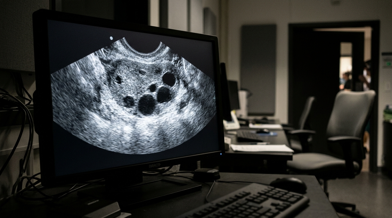
The new system, called MSERNet, was trained and tested on a private, ethically approved collection of retrospective ovarian ultrasound scans and on an independent public carotid artery dataset, and its results are reported in the journal Frontiers in Artificial Intelligence.
The problem the researchers tackled is subtle but consequential. Ultrasound is prized in routine clinical practice because it offers real-time imaging, portability, safety, and low cost compared with computed tomography and magnetic resonance imaging. Yet the irreversible compression that makes digital storage practical can introduce blocking distortion, blurring, and loss of the speckle texture that carries diagnostic information. Excessive compression can therefore affect both visual interpretation and the performance of automated image analysis. While deep learning methods for removing JPEG artifacts have surged recently, the authors note that few studies have addressed compression-aware restoration on clinically realistic datasets with well-controlled degradation strategies, and problems such as consistent preprocessing, photometric standardization, and reliable lossless ground-truth data remain unresolved.
MSERNet’s architecture combines four complementary modules within a single residual learning framework. An initial feature extraction block of two consecutive 3 × 3 convolution layers with ReLU activation pulls out low-level image features. A multi-scale block then runs parallel 3 × 3, 5 × 5, and 7 × 7 convolutions to capture information at different receptive fields, and the resulting feature maps are concatenated and fused through a 1 × 1 convolution. An adaptive feature fusion module applies channel attention using global average pooling to emphasize informative responses and suppress less relevant ones. An edge refinement module, built from two 3 × 3 convolution layers with a residual connection, sharpens structural boundaries. Finally, a residual prediction layer estimates the compression-artifact residual, and the restored image is produced by subtracting that estimated residual from the compressed input. This residual strategy lets the network concentrate on artifact removal while leaving the original image content untouched.
The dataset work was deliberately careful. The private dataset contained 546 anonymized ovarian ultrasound DICOM images from ES Hospital in India, collected between January and December 2024 during routine clinical examinations, with one image selected from each patient-coded folder so that no patient appeared in more than one subset. The study was approved by the Institutional Ethics Committee of Vellore Institute of Technology, which waived written informed consent because the data were retrospective and fully anonymized. A second dataset, the Common Carotid Artery ultrasound collection from Mendeley Data, contributed 546 randomly selected public images. All images were converted to grayscale and saved as lossless PNG files at a uniform resolution of 468 × 650 pixels, giving the restoration framework a clean reference standard that introduces no compression artifacts of its own.
Degradation was simulated under controlled conditions. Each reference image was compressed at four JPEG quality factors—Q30, Q50, Q70, and Q85—with lower quality factors producing more visible artifacts. Two repeated compression scenarios, Q70 followed by Q30 and Q85 followed by Q50, were also generated to mimic images that get compressed multiple times during storage, transmission, or archiving. The 546 source images were split into mutually exclusive training, validation, and test subsets of 382, 82, and 82 images using a fixed random seed, and all degradation versions of the same source image stayed within the same subset to prevent patient and source-image leakage. Training used 256 × 256 patches, a batch size of 8, the Adam optimizer with an initial learning rate of 1 × 10⁻⁴, and an L1 loss, with a ReduceLROnPlateau scheduler adjusting the learning rate during 100 epochs on an NVIDIA Tesla T4 GPU. The final model has just 466,641 trainable parameters, roughly 0.467 million.
Performance was measured with three standard image quality metrics: Peak Signal-to-Noise Ratio, which gauges pixel-level similarity; the Structural Similarity Index, which evaluates luminance, contrast, and structure; and Visual Information Fidelity, which quantifies how much visual information survives restoration. On the private ovarian dataset, MSERNet reached a PSNR of 47.06 ± 1.19 dB, SSIM of 0.9904 ± 0.0030, and VIF of 0.8026 ± 0.0551 at Q85, and still achieved 39.33 ± 0.96 dB PSNR at the harshest Q30 setting. Recompressed images followed the same pattern, with Q70_Q30 yielding 39.07 ± 0.96 dB and Q85_Q50 yielding 42.73 ± 1.02 dB. On the public carotid dataset the numbers were higher still, peaking at 48.93 ± 1.28 dB PSNR, 0.9954 SSIM, and 0.8916 VIF at Q85, and the training curves on both datasets converged smoothly with no clear signs of overfitting.
Cross-dataset tests probed generalization without any additional fine-tuning. A model trained only on the private ovarian images and tested directly on the public carotid images achieved 48.53 ± 1.21 dB PSNR at Q85 and 46.05 ± 1.19 dB at Q70. The reverse experiment—training on public carotid images and testing on private ovarian scans—produced 42.80 ± 1.38 dB at Q85 and 38.93 ± 1.04 dB at Q30. A paired t-test on the 82-image public test set compared the public-domain model with the cross-domain model image by image and found the public-domain evaluation consistently ahead, with differences ranging from 0.40 to 1.68 dB in PSNR, all statistically significant at p < 0.001. The authors explicitly caution that statistical significance was not interpreted as evidence of clinical equivalence.
Head-to-head comparisons against established restoration methods revealed a nuanced picture. MSERNet posted the highest PSNR at Q50, Q70, Q85, and Q85_Q50, reaching 47.06 ± 1.19 dB at Q85, while the DCT-domain spatial-frequential Transformer of Ouyang and Chen achieved higher PSNR at Q30 and Q70_Q30. For SSIM, MSERNet led at Q50 and Q85, whereas PALMSNet recorded higher values under other conditions, and for VIF, MSERNet topped Q70 and Q85 while Wiener filtering led elsewhere. No single method dominated every metric and degradation level, which the authors say underscores that restoration behavior depends on both the compression condition and the restoration approach.
Efficiency and downstream utility rounded out the evaluation. Five repeated training runs with different random seeds produced PSNR values ranging from 39.00 ± 1.01 to 46.03 ± 1.28 dB across the six degradation conditions, indicating the measured performance was not dependent on a single run. Computationally, MSERNet’s 0.467 million parameters and 1.78 MB model size were far smaller than RRDB’s 1.906 million parameters and 22.01 MB or PALMSNet’s 7.417 million parameters and 84.98 MB, and its 141.17 giga-MACs and 71.20 millisecond inference time per image undercut both, though the lighter ARCNN remained cheaper across all measures.
The authors are careful about what these results do and do not establish. The evaluation used grayscale images from two datasets under JPEG compression only, and other imaging modalities and compression standards were not included. The downstream classification relied on a 53-image test set and does not directly establish improved clinical diagnostic performance. Visual comparisons showed reduced blocking artifacts, clearer lesion boundaries, and minimal over-smoothing on both datasets and in cross-dataset tests, consistent with the quantitative gains, but the metrics alone cannot confirm a diagnostic benefit. Future work, the team writes, will evaluate MSERNet on larger and more diverse datasets, additional compression schemes, and other medical image-analysis applications including lesion segmentation, detection, and classification.
Subject of Research: Deep learning restoration of JPEG compression artifacts in retrospective ovarian ultrasound images
Article Title: MSERNet: a multi-scale edge-guided network for JPEG compression artifact restoration in retrospective ovarian ultrasound images
Article References: Komala, K., & Kalaivani, S. (2026). MSERNet: a multi-scale edge-guided network for JPEG compression artifact restoration in retrospective ovarian ultrasound images. Frontiers in Artificial Intelligence, 9, Article 1964967. https://doi.org/10.3389/frai.2026.1964967
Image Credits: AI Generated
DOI: 10.3389/frai.2026.1964967
Keywords: ultrasound, JPEG compression, artifact restoration, deep learning, ovarian imaging, residual learning, multi-scale feature extraction, image quality metrics, cross-dataset evaluation, transfer learning, medical image analysis, MSERNet
News Source: Blake Davidson. (November 6, 2026). Deep learning network strips JPEG artifacts from ovarian ultrasound images. Scienmag.