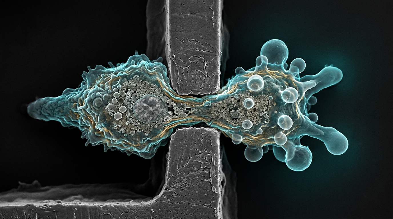
Social amoebas squeezing beneath a layer of agarose have revealed a new job for a familiar molecular scaffold. When Dictyostelium discoideum cells lacking PaxB, the amoeba’s version of the mammalian adhesion protein paxillin, were pushed into ever more confined spaces, they produced blebs that were disproportionately large relative to their body size, while normal cells kept their bleb-to-cell proportions steady. The finding, published in BMC Molecular and Cell Biology, pairs live-cell imaging with a one-dimensional mechanical model of bleb expansion to argue that PaxB helps control how quickly a new actin cortex assembles beneath a freshly formed bleb, and that this assembly rate, not pressure alone, sets the final size of the protrusion.
Blebs are blister-like bulges that cells throw out when the membrane locally tears away from the actin cortex beneath it and is shoved outward by intracellular fluid pressure. Unlike pseudopods, which advance by actin polymerization at the leading edge, blebs dominate movement in three-dimensional, confined settings such as the spaces between cells in a tissue or the pores of an extracellular matrix. Cancer cells, immune cells and Dictyostelium all use them. Yet the molecular rules governing when a bleb starts, how big it gets and how it stabilizes remain incompletely mapped, which is precisely the gap the new study set out to narrow.
Paxillin, in mammals, sits at focal adhesions where it links integrins to the actin cytoskeleton and recruits signaling molecules that coordinate adhesion, cytoskeletal remodeling and migration. Dictyostelium carries a paxillin ortholog, PaxB, that shares conserved domains with its mammalian counterpart and participates in adhesion, cytokinesis, development and chemotaxis. Whether paxillin-family proteins contribute to bleb-based motility, however, had been unclear. Intriguingly, earlier work in mammalian systems suggested that losing paxillin can enhance invasion in three-dimensional environments, hinting that the protein might influence bleb formation or dynamics in some way.
To test that idea, the researchers grew wild-type Ax2 cells and paxB knockout cells engineered to express LifeAct-GFP, a fluorescent tag that labels filamentous actin, and coaxed them to crawl under agarose gels laced with rhodamine-dextran toward a source of cyclic AMP, the amoeba’s chemoattractant. Two gel concentrations, 0.4 percent and 0.7 percent, imposed low and high compression respectively. A spinning-disc confocal microscope captured the membrane position against the dextran background and the cortex in green, at roughly 1.7 to 1.9 seconds per frame for 30-second windows. Blebs were scored when the membrane visibly separated from the cortex and a new cortex reformed beneath it, and their cross-sectional areas were measured from the first frame in which the bleb reached a stable size.
The first result was a null: bleb frequency was essentially identical between the two strains. Under low compression, wild-type cells averaged 4.4 blebs per cell over the observation window while paxB knockouts averaged 4.2; under high compression the figures rose to 8.25 and 7.89 respectively, with no statistically significant difference between genotypes in either condition. Both cell types did bleb significantly more often when squeezed harder, confirming that compression drives blebbing frequency regardless of PaxB status. Whatever PaxB does, the data suggest, it is not about how many blebs a cell launches.
Size told a different story. Absolute bleb area was larger in wild-type cells than in knockouts under both compression levels, but the percentage increase in bleb size when compression rose was nearly the same in both strains, roughly 73 percent for Ax2 and 68 percent for paxB knockouts. The surprise came when the researchers normalized bleb area to cell cross-sectional area, a metric they call relative bleb size. Wild-type cells, which flattened dramatically under the heavier gel, expanding their cross-sectional area by about 90 percent, held their relative bleb size essentially constant, shifting only about 6 percent. The knockouts, whose cell area grew only about 10 percent under the same load, boosted their relative bleb size by 52 percent, from 0.0158 to 0.0244. Because cell flattening under load is associated with rising hydrostatic pressure, the modest area change in the knockouts implies their internal pressure did not climb much, yet their blebs still swelled out of proportion.
That combination pointed the investigators toward cortex assembly rather than pressure as the lever PaxB might pull. Bleb formation is a cycle: the membrane detaches, fluid floods the blister, and then a new actin cortex reassembles beneath the protruded membrane while the old cortex, sometimes called an actin scar, is degraded. Paxillin is known to regulate actin-based processes, and prior work had found that PaxB, along with phospholipase D, governs actin polymerization in Dictyostelium. If PaxB loss slowed or mistimed the assembly of the new cortex, the bleb would spend more of its expansion phase pushing against a flimsier boundary and could settle at a larger final size even without extra pressure.
To turn that hypothesis into something testable, the team built a one-dimensional mechanical model of bleb expansion. The cell boundary is treated as a composite of an elastic membrane and a viscoelastic cortex, with linker proteins whose force-dependent binding and unbinding resist membrane separation. Cytoplasm is modeled as a viscous fluid coupled in parallel with the boundary, and the driving force is an exponentially decaying pressure that starts high at nucleation and falls as the bleb grows. Crucially, the model includes time-dependent stiffness and viscosity terms tied to the kinetics of actin polymerization and depolymerization during cortex reformation and degradation, parameters drawn from the group’s earlier data-driven work on bleb stabilization. Solving the equations with MATLAB’s stiff ODE solver, the researchers first validated the model against known bleb behavior: simulated blebs grew with intracellular pressure, shrank with stiffer membranes and cortices, and showed the partial retraction toward equilibrium size that experiments have documented as new cortex assembles.
An equilibrium analysis of the model then delivered the key theoretical insight. The steady-state bleb size depends on initial pressure, membrane stiffness, cortex stiffness and, notably, the ratio of actin polymerization to depolymerization rates in the reforming cortex, but not on cortex degradation rate, cytoplasmic viscosity or cortex permeability. Lowering that polymerization ratio yields larger blebs at the same pressure. When the team simulated cells flattening under increasing compression, calibrated against measured cell heights and areas, the wild-type scenario reproduced the observed 6 percent shift in relative bleb size. For the knockout, holding the 10 percent area increase fixed and dialing the polymerization ratio down from the wild-type value of 1.75 to between 1.094 and 1.313 reproduced the 52 percent jump in relative bleb size.
The authors are careful about the model’s limits. The one-dimensional formulation uses a phenomenological exponential decay for intracellular pressure and ignores lateral membrane tension and three-dimensional surface curvature, simplifications they argue do not overturn the central conclusion because the study’s focus is cortex assembly rather than bleb shape. Their pressure estimates via the Young-Laplace equation also neglect the viscoelastic damping of the cytoplasm, so absolute pressures may be overestimated, though the conclusions rest on pressure changes rather than absolute values. Future experiments, they write, will directly quantify actin polymerization and depolymerization rates in the reforming bleb cortex of paxB knockout cells to validate the hypothesized mechanism.
Subject of Research: Role of the paxillin ortholog PaxB in regulating bleb size during confined cell migration in Dictyostelium discoideum
Article Title: A role for PaxB in regulating blebbing: experimental insights and theoretical perspectives from Dictyostelium discoideum
Article References: Santiago, Z., Wey, B., Sewwandi de Silva, S. H. D., Reznik, J., Siby, L., Asante-Asamani, E., & Brazil, D. (2026). A role for PaxB in regulating blebbing: experimental insights and theoretical perspectives from Dictyostelium discoideum. BMC Molecular and Cell Biology, 27(1), Article 25. https://doi.org/10.1186/s12860-026-00584-w
Image Credits: AI Generated
DOI: 10.1186/s12860-026-00584-w
Keywords: PaxB, paxillin, blebbing, Dictyostelium discoideum, cell migration, chemotaxis, actin cortex, cortex reformation, mathematical modeling, under-agarose assay, cell compression, hydrostatic pressure
News Source: Drew Townsend. (November 6, 2026). PaxB keeps blebs in check as cells squeeze through tight spaces. Scienmag.