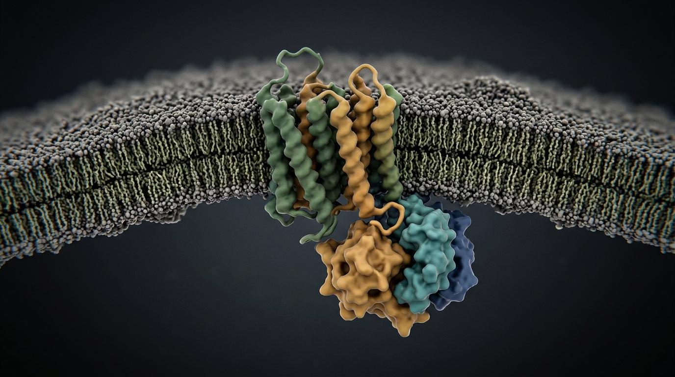
Heterotrimeric G proteins act as molecular switches that relay signals from G-protein-coupled receptors into cells, and their activation has long been attributed mainly to the C-terminal α5 helix of the Gα subunit, the region that grips the receptor. A study published in Nature Chemical Biology now shows that a second structural element, the N-terminal helix of Gαs, actively promotes the release of GDP that switches the protein on. Using single-molecule diffraction tracking, molecular dynamics simulations and engineered crosslinks, the researchers found that a counterclockwise rotation of this helix, viewed from the extracellular side, loosens the nucleotide-binding pocket, whereas clockwise rotation does not.
The team, led by Hiroki Arai and Asuka Inoue of Tohoku University with colleagues in Japan, the United States and Canada, used diffracted X-ray tracking to watch the helix move in real time. GDP-bound Gs heterotrimer was immobilized on a surface through a tag on the Gγ subunit, and gold nanocrystals were attached to surface-exposed methionine and cysteine residues concentrated at the tip of the N-terminal helix. Purified β3-adrenergic receptor in detergent micelles was then added, and the sample was irradiated with 0.11-nanometer X-rays at 100 microseconds per frame at the SPring-8 synchrotron. The angular movement of each diffraction spot was decomposed into two components: θ, rotation within the plane of the substrate, and χ, rotation about an axis perpendicular to it.
Across three conditions—no receptor, unliganded receptor and agonist-bound receptor—the θ component showed no appreciable differences. The χ component, however, displayed a marked increase in mean squared displacement only when the agonist isoproterenol was bound to the receptor, indicating that engagement with an active GPCR selectively enhances rotational fluctuations of the helix on a submillisecond timescale. Because the Debye–Stokes–Einstein relation ties rotational diffusion inversely to molecular volume, the selective enhancement along one axis could not be explained by the receptor simply adding mass. The differences were most pronounced at early time points of 100 to 200 microseconds, and analysis of step sizes showed that the active receptor increased the probability of large angular displacements of roughly 3 to 10 milliradians in both positive and negative directions, a bidirectional pattern the authors attribute to continuous association and dissociation of the receptor–G protein complex under GDP-bound conditions.
A complementary diffracted X-ray blinking analysis confirmed that the fast-moving signals came from the helix itself. A double mutant in which the two primary labeling sites were replaced showed roughly half the diffraction intensity of the wild type, and the fast-mobility population seen in the wild type was markedly reduced in the mutant. The data therefore support the conclusion that the rapid dynamics detected in the tracking experiments primarily reflect motion of gold nanocrystals attached to the N-terminal helix, although the tracking data alone could not determine the direction of rotation.
To resolve direction, the researchers turned to structural statistics and simulation. From 377 experimentally determined structures of active-state GPCR–Gs complexes, they assembled a nonredundant set of 53 distinct complexes, aligned them by the Ras homology domain of Gα, and defined an axis through the base of the N-terminal helix using the inactive structure as reference. Viewed along this axis, the tip residue of the helix sat toward the upper left in every active structure relative to inactive Gs, consistent with a rotational displacement. In the interaction network, the G.hns1 region at the base of the helix contacted the receptor in more than half of the complexes, and an interaction involving the residues Arg38 and Ala39 was present in roughly 60 percent of them, with a positively charged residue conserved at these positions across Gα subfamilies.
Functional tests with a NanoBiT split-luciferase assay that monitors Gα–Gβ dissociation showed that replacing Arg38 with alanine lowered activation for four of five Gs-coupled receptors tested—β3AR, β2AR, the dopamine D1 receptor and the 5-HT4 receptor—while substituting arginine at position 39 elevated dissociation responses across all five, which also included the vasopressin V2 receptor. A separate assay measuring GPCR–Gα association showed that the mutations left the association step largely intact, indicating that the effects arose at the dissociation stage rather than at initial receptor engagement. Structural inspection of the interface between the receptor’s intracellular loop 2 and the helix explained some, though not all, of the receptor-dependent differences, and the authors suggest the helix may act during an intermediate state of complex formation that current structures have not captured.
The decisive evidence for direction came from molecular dynamics. Simulating a membrane-embedded β2AR–Gs complex in a lipid bilayer, the team observed nucleotide dissociation in one of four 900-nanosecond production runs. In that trajectory, the α5 helix straightened at about 15 nanoseconds, the α-helical domain opened at about 430 nanoseconds, and GMP, a lower-affinity stand-in for GDP, dissociated at about 700 nanoseconds. Between roughly 400 and 550 nanoseconds, before nucleotide release, the N-terminal helix gradually underwent the counterclockwise displacement, in concert with receptor intracellular loop engagement, domain opening and dissociation. Restrained simulations then tested causality: when the helix was forced to rotate counterclockwise while a restraint pulled the β6–α5 region toward the active conformation, GDP displacement increased in six of ten simulations, whereas clockwise restraint produced no such effect. Without the β6–α5 restraint, GDP release did not occur under either condition, showing that the two pathways cooperate.
Biochemical crosslinking validated the simulations. The researchers engineered Gαs mutants with paired cysteines designed to lock the helix in either the counterclockwise or clockwise position, crosslinked them with a cleavable reagent, and measured nucleotide exchange with a fluorescent GDP analog in POPC nanodiscs. The counterclockwise-locked protein showed enhanced GDP exchange and release that was abolished when the crosslink was chemically reduced, while the clockwise-locked protein showed no change. The same result held for lipidation-free protein purified from bacteria, demonstrating that the counterclockwise effect operates even without a membrane.
The pathway’s route through the protein emerged from analysis of the β-sheet network. In the simulations that achieved release, residues on the αG helix that contact the guanine ring of GDP flipped outward, and rigidity transmission allostery analysis traced signal flow from the hinge between the N-terminal helix and the β1 strand through the β4 and β5 sheets to the αG helix and the P-loop, with transmission patterns that depended on the direction of helix motion. Counterclockwise rotation twisted β1, widened the gap between β1 and β4, narrowed the gap between β4 and β5, and reorganized the hydrophobic side-chain network connecting the sheets. The authors propose that this allosteric route operates alongside the established α5-mediated pathway: the receptor first pulls α5 upward, loosening the sheet network, after which the helix contacts the receptor’s intracellular loops and rotates counterclockwise, transmitting structural change to the guanine-binding site and the P-loop. Because the helix and the sheet residues are conserved across Gα subtypes, the researchers suggest this mechanism may underlie nucleotide release throughout the G-protein family, though distinguishing between receptor-driven and domain-opening contributions to the rotation will require methods with higher spatiotemporal resolution.
Subject of Research: Rotational dynamics of the Gαs N-terminal helix in GPCR-triggered G protein activation
Article Title: Rotation of the N-terminal helix triggers Gα activation
Article References: Arai, H., Ikuta, T., Kise, R., Tatsumi, M., Koizumi, Y., Yanagawa, M., Sasaki, D., Tawfeeq, C., Oshima, H. S., Yau, W.-I., Shihoya, W., Sljoka, A., Taninaka, K., Nureki, O., Sunahara, R. K., Mio, K., Sasaki, Y. C., & Inoue, A. (2026). Rotation of the N-terminal helix triggers Gα activation. Nature Chemical Biology. https://doi.org/10.1038/s41589-026-02359-y
Image Credits: AI Generated
DOI: 10.1038/s41589-026-02359-y
Keywords: GPCR, G protein, Gαs, N-terminal helix, GDP release, diffracted X-ray tracking, molecular dynamics, allosteric signaling, β-sheet network, crosslinking, β3-adrenergic receptor, conformational change
News Source: Drew Townsend. (November 5, 2026). N-terminal helix rotation drives GPCR-triggered G protein activation. Scienmag.