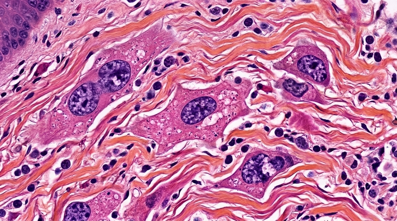
Skin that refuses to heal, skin that stays inflamed for months, and skin that relapses despite treatment may share a hidden driver: senescent cells. A wide-ranging review published in Frontiers in Aging argues that cellular senescence, the stable state in which damaged cells stop dividing but remain metabolically active, could be a mechanism linking chronic tissue stress to persistent skin inflammation. The authors, dermatology researchers at Shanghai University of Traditional Chinese Medicine, systematically map how senescent cells sense stress, secrete inflammatory molecules, evade immune clearance, and shape disease outcomes across a spectrum of inflammatory skin conditions.
Senescent cells are not simply old cells. The review describes them as cells locked into stable cell-cycle arrest by diverse endogenous and exogenous stressors, accompanied by characteristic changes in morphology, metabolism, and secretory behavior. The central feature is the senescence-associated secretory phenotype, or SASP, through which these cells release proinflammatory cytokines, chemokines, matrix metalloproteinases, and extracellular vesicles into the surrounding microenvironment. SASP can act locally or systemically, recruiting immune cells, remodeling the extracellular matrix, and even inducing senescence in neighboring cells, creating a self-amplifying loop of tissue inflammation.
What makes cutaneous senescence difficult to study is that no single marker is sufficient to identify it. The p16INK4a protein may appear in non-senescent or differentiated cells, p21CIP1 can indicate a reversible checkpoint response rather than permanent arrest, and SA-β-gal activity reflects lysosomal expansion that also occurs in quiescent cells or activated macrophages. Low Ki-67 alone cannot distinguish senescence from quiescence or terminal differentiation. The review therefore insists that senescence in skin requires multiparameter, cell-type-resolved criteria, combining cell-cycle arrest markers with auxiliary hallmarks and spatial localization, rather than imposing a universal numerical threshold.
The triggers of senescence in skin are layered. Endogenous factors include oxidative stress from mitochondrial reactive oxygen species, telomere shortening with repeated cell division, persistent DNA damage, proteostasis disruption, and carbonyl stress from advanced glycation end products. Exogenous factors include ultraviolet radiation, particulate pollution, and ionizing radiation. These stressors converge on shared molecular nodes: the p16INK4a/pRB and p53/p21CIP1 tumor-suppressive pathways regulate the arrest program, while persistent DNA damage activates cGAS-STING-dependent inflammatory signaling that links damage sensing directly to SASP regulation.
Not all senescence is harmful. The review emphasizes that short-term, localized SASP can participate in tissue repair. Senescent-cell-derived PDGF-AA is required for normal wound healing, and transient SASP exposure can enhance keratinocyte proliferative capacity. Senescence and SASP are therefore not inherently pathological. The problem arises when senescent cells persist or fail to be cleared, shifting the same secretory program from a repair signal toward chronic proteolysis, inflammatory amplification, and paracrine senescence that spreads the damage signal to surrounding healthy tissue.
The strongest evidence for senescence as a disease driver comes from chronic and diabetic wounds. Human chronic wounds and diabetic foot ulcers show increased p16 and p21 expression, senescence-gene and SASP signatures, and an association between higher p21 levels and delayed healing. In a cohort of 79 patients with chronic wounds, Cox modeling showed that high CDKN1A expression predicted prolonged healing time. Experimental studies identify dermal fibroblasts and persistent SASP-producing macrophages as candidate effectors, and pharmacological clearance of senescent cells improves wound repair in animal models. DPP4-targeted nanospheres can selectively eliminate senescent fibroblasts in diabetic wounds, downregulate SASP, induce M2 macrophage polarization, and accelerate re-epithelialization and collagen deposition.
In other inflammatory skin diseases, the evidence is more associative. Psoriasis shows lesional keratinocytes and CD4+ T cells with senescence-associated phenotypes, and preclinical interventions such as topical ABT-737 and CD4-specific Tet2 deletion improve psoriasiform dermatitis in mice, but human senescence-selective trials are absent. Atopic dermatitis evidence is largely restricted to elderly patients, where fibroblast- and SASP-enriched programs are associated with macrophage and NK-cell recruitment. Cutaneous lupus erythematosus has a comparatively coherent chain, with human p21-high basal epidermal cells linked to type I interferon amplification and fisetin or topical ABT-737 improving lupus-related skin disease in mice.
The review classifies senescence-targeted interventions into three categories. Senolytics selectively induce senescent cell death by exploiting their dependence on anti-apoptotic pathways. Dasatinib plus quercetin selectively cleared senescent fibroblasts in a human-mouse skin chimera model, and dasatinib monotherapy reduced senescent-cell burden and fibrosis in human keloid tissue. Navitoclax and ABT-737 induce apoptosis of senescent fibroblasts in vitro and clear UV-induced senescent dermal fibroblasts in mouse skin. Senomorphics aim to modify the secretory phenotype of already senescent cells without killing them. Rapamycin currently has relatively clear human-skin senomorphic evidence, with topical treatment reducing epidermal p16INK4a expression and increasing collagen VII content in aged skin. Senopreventive or geroprotective strategies, including metformin, hesperidin, and magnolin, aim to prevent the establishment of senescence before or during stress exposure.
The review also highlights the therapeutic paradox of phototherapy. Narrowband UVB can induce apoptosis of T cells in psoriatic lesions, and a randomized controlled trial showed that NB-UVB improved disease activity in adults with moderate-to-severe atopic eczema compared with a visible-light control. Yet clinical improvement alone does not demonstrate senolysis or senomorphic activity; such claims require multiparameter identification of senescent target cells and evidence of selective clearance or SASP modulation. The authors caution that without cellular source and senescent-state evidence, established immunomodulatory drugs should still be regarded as anti-inflammatory therapies rather than being directly equated with senescence-targeted drugs.
Translating cellular senescence into a therapeutic target requires more than documenting marker-positive cells. The authors call for defining senescence-maintained lesion subtypes, establishing combinatorial biomarkers and spatial maps, and validating the efficacy and safety window of local therapies in suitable models and human tissues. Mouse skin differs from human skin in immune composition, tissue architecture, and inflammatory chronicity, and senotherapeutics often act only on senescent states in specific cell types. The safety window will also determine translational feasibility: rapamycin can suppress general immune function, dasatinib can affect hematopoiesis, and BCL-2/BCL-XL inhibitors carry hematological toxicity risks. Local delivery through topical treatment, intradermal injection, microneedles, and responsive nanocarriers may limit systemic exposure and move senescence-targeted therapy from a systemic anti-aging concept toward precision treatment of defined cutaneous lesions. If future studies demonstrate that altering senescent cells or their secretory programs within lesions improves inflammation and promotes matrix repair, senescence-targeted therapy may become an important adjunct to existing immunomodulatory approaches.
Subject of Research: The role of cellular senescence and the senescence-associated secretory phenotype in driving chronic inflammatory skin disease and the potential of senolytic and senomorphic therapies.
Article Title: Cellular senescence in skin inflammation: molecular mechanisms and potential clinical implications
Article References: Wang, X., Xiong, B., Li, S., Wang, Y., & Li, F. (2026). Cellular senescence in skin inflammation: molecular mechanisms and potential clinical implications. Frontiers in Aging, 7, Article 1986107. https://doi.org/10.3389/fragi.2026.1986107
Image Credits: AI Generated
DOI: 10.3389/fragi.2026.1986107
Keywords: cellular senescence, SASP, skin inflammation, senolytics, senomorphics, chronic wounds, diabetic foot ulcers, psoriasis, atopic dermatitis, cutaneous lupus erythematosus, systemic sclerosis, vitiligo
News Source: Ophelia Keating. (November 5, 2026). Senescent Cells May Hold the Key to Chronic Skin Inflammation, Review Finds. Scienmag.