


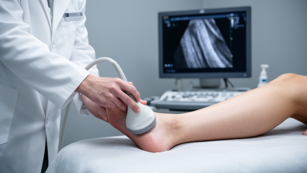
People with type 2 diabetes who also carry a diagnosis of peripheral nerve damage show measurably thicker and larger Achilles tendons than diabetic patients whose nerves remain intact, according to a new ultrasonographic study published in Egyptian Rheumatology and Rehabilitation. The research, led by Youssy Saeed Eshak Gergius and colleagues, compared 52 patients with type 2 diabetes recruited from the Diabetes outpatient clinic of the Endocrinology department at a university hospital, splitting them evenly into a group with diabetic peripheral neuropathy and a group without it. Every participant underwent clinical assessment, laboratory testing, nerve conduction studies, and a standardized musculoskeletal ultrasound examination of both Achilles tendons, allowing the team to compare ultrasonographic tendon abnormalities between patients with and without diabetic peripheral neuropathy.
Patients with diabetic peripheral neuropathy had significantly greater Achilles tendon thickness and significantly larger cross-sectional area on both the right and left sides, with p values at or below 0.001 for these comparisons. The tendon’s internal texture told a similar story: an abnormal fibrillar pattern, in which the normally organized linear echoes of healthy collagen are disrupted, appeared far more often in the neuropathy group, reaching statistical significance on the right side at p = 0.006 and on the left side at p = 0.001.
Recruitment for the study took place over ten months, during which 62 patients with type 2 diabetes were initially enrolled. Seven were excluded for not meeting the full inclusion criteria and three declined to complete all required investigations, leaving 52 participants, 26 in each arm. The two groups were matched for age and sex, and all participants were between 18 and 65 years old with no musculoskeletal symptoms in the lower limb joints, meaning the tendon changes were detected in people who had no pain, swelling, stiffness, or other clinical signs of Achilles tendinopathy. Patients with traumatic nerve injury, lumbar radiculopathy, foot or ankle fractures, foot ulcers, Charcot neuroarthropathy, smoking or alcohol histories, systemic disorders causing polyneuropathy, prior Achilles tendon injuries, rheumatological diseases, athletic training backgrounds, congenital ankle deformities, or chronic steroid use were all screened out.
By this scheme, eight patients were classified as mild, four as moderate, and fourteen as severe.
The severity analysis suggested a dose-like pattern. When the researchers compared tendon measurements across the three severity categories, statistically significant differences emerged in Achilles tendon thickness on both sides, with p values of 0.012 and 0.036, and in cross-sectional area on the left side, with p = 0.023. Post hoc Tukey testing showed that the significant differences occurred primarily between the mild and severe groups, and abnormal fibrillar patterns were present in roughly two-thirds of patients with severe neuropathy. Neither retrocalcaneal bursitis nor enthesophytes, the bony projections at the tendon’s insertion, differed significantly across severity groups, and no intratendinous Doppler signal was detected in either study group.
DPN status emerged as the strongest independent predictor of both thickness and cross-sectional area on both sides, with standardized coefficients ranging from 0.457 to 0.501 and p values at or below 0.002. Diabetes duration also contributed significantly in all four models, while cholesterol reached significance only for the left-sided measurements. Binary logistic regression, again adjusted for duration and cholesterol, showed that patients with neuropathy had approximately fivefold higher odds of an abnormal fibrillar pattern on the right side and twenty-two-fold higher odds on the left side, with p values of 0.044 and 0.010 respectively. Neither diabetes duration nor cholesterol independently predicted fibrillar abnormalities after adjustment.
The authors interpret these findings through the biology of diabetic tissue injury. Chronic hyperglycemia drives the accumulation of advanced glycation end products, which provoke excessive collagen cross-linking and impair collagen turnover, reducing tendon elasticity and repair capacity. Diabetic microangiopathy may further compromise the tendon’s blood supply, while loss of sensory innervation could add denervation-related trophic changes, impaired proprioception, and abnormal loading patterns during gait, collectively accelerating tendon remodeling. Experimental evidence cited in the paper indicates that peripheral nerves play a crucial role in tendon maintenance, repair, and mechanotransduction, with neural-derived mediators regulating collagen synthesis, vascular responses, and tissue remodeling, so the loss of neural input may directly undermine tendon homeostasis.
Previous studies have shown Achilles tendon thickening and stiffness in diabetes, changes that were found to lead to a reduction in ankle joint range of motion, which in turn may alter stance-phase biomechanics and increase forefoot loading, with excessive weight-bearing stress beneath the metatarsal heads potentially predisposing patients to a greater risk of foot ulcers. Because the tendon changes in this study were found in asymptomatic patients, the authors conclude that musculoskeletal ultrasound is a valuable tool for the early detection of subclinical tendon involvement in patients with type 2 diabetes and diabetic peripheral neuropathy. The authors suggest that patients with diabetic peripheral neuropathy may benefit from interventions that reduce tendon loading, noting that appropriate footwear, silicone insoles, and stretching exercise, together with control of diabetes, have been previously proposed, although their effectiveness was not evaluated in this study.
Important caveats accompany the results. The cross-sectional design cannot establish that neuropathy causes the tendon changes, only that the two are associated. The study was conducted at a single center with a modest sample size, and the moderate neuropathy subgroup contained only four patients, limiting statistical power for the severity comparisons. The regression analyses were exploratory and carry an elevated risk of overfitting, with wide confidence intervals in some estimates. Healthy non-diabetic controls were not included, so the findings distinguish diabetic patients with neuropathy from those without but cannot isolate diabetes itself as the source of the changes. Factors such as long-term glycemic control, physical activity, occupational loading, foot biomechanics, and medications including statins were not systematically assessed, and formal intra-observer reliability statistics were not calculated. Elastographic assessment of tendon stiffness was also not performed.
The authors call for prospective longitudinal studies in larger cohorts to determine whether ultrasonographic Achilles tendon abnormalities predict clinically important outcomes such as tendon rupture, gait impairment, diabetic foot ulceration, and long-term functional disability.
Subject of Research: Ultrasonographic changes of the Achilles tendon in type 2 diabetes patients with and without diabetic peripheral neuropathy
Article Title: Effect of peripheral neuropathy on Achilles tendon: ultrasonographic study
Article References: Gergius, Y. S. E., Elnaby, M. M. H., Motaher, A. M. F. M., & Moussa, S. G. (2026). Effect of peripheral neuropathy on Achilles tendon: ultrasonographic study. Egyptian Rheumatology and Rehabilitation, 53(1), Article 72. https://doi.org/10.1186/s43166-026-00445-6
Image Credits: AI Generated
DOI: 10.1186/s43166-026-00445-6
Keywords: diabetic peripheral neuropathy, Achilles tendon, type 2 diabetes, musculoskeletal ultrasound, tendon thickness, cross-sectional area, fibrillar pattern, nerve conduction studies, tendon degeneration, advanced glycation end products, diabetic foot complications, cross-sectional study
News Source: Ophelia Keating. (October 30, 2026). Nerve Damage Reshapes the Achilles Tendon in Diabetes, Ultrasound Study Finds. Scienmag.



