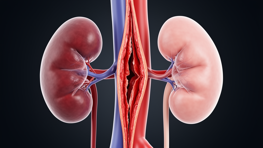
Kidneys sometimes shrink after surgery for aortic dissection even when the renal artery looks perfectly open on follow-up imaging. A new exploratory study used patient-specific computational fluid dynamics (CFD), built from serial computed tomography angiography (CTA) scans, to compare blood-flow conditions around the renal arteries in patients whose kidneys later atrophied with those in patients whose kidneys kept their volume. The researchers report that two measures of false-lumen flow near the renal arteries were lower in every kidney that later atrophied than in every comparator kidney, beginning in the early postoperative period.
The study, published in Frontiers in Bioengineering and Biotechnology, focused on six index kidneys from six men: three with at least 30% loss of reference kidney volume and three age-matched comparators. All six kidneys were supplied by the false lumen before repair. The team built CFD models from preoperative, early postoperative and late follow-up CTAs, keeping inlet and outlet boundary conditions identical across time points within each patient so that changes could be attributed to the evolving geometry rather than to differing model assumptions.
Aortic dissection creates two channels: the true lumen, through which blood normally flows, and a false lumen within the vessel wall. The two channels communicate through tears, and the behavior of flow through these communications is thought to influence how branches such as the renal arteries are perfused. In total, the researchers characterized 90 true-lumen/false-lumen communications by their distance from the renal-ostial plane, their direction, the pressure difference across them and the trans-tear flow. They also quantified true- and false-lumen relationships at the renal-ostial plane and computed bilateral renal-artery flow directly from the models. Kidney volume was measured by three-dimensional CTA segmentation.
The volume outcomes were stark. Index-kidney volume changed by −53.0%, −64.1% and −50.6% in the atrophy group, compared with −13.8%, +0.8% and −10.7% in the comparators. Yet before repair, no hemodynamic measure separated the two groups, suggesting that the discriminating features emerged only after the operation altered the dissection’s flow dynamics.
From the early postoperative model onward, at 3 to 37 days after surgery, two aortic-level flow measures distinguished every atrophy kidney from every comparator. The false-lumen flow-magnitude fraction at the renal-ostial plane ranged from 0.03 to 0.10 in the atrophy group versus 0.13 to 0.79 in comparators, and total peak-systolic trans-communication flow ranged from 37.8 to 45.1 mL/s versus 61.8 to 185.3 mL/s. In other words, kidneys that later shrank were surrounded by less false-lumen flow activity near the renal arteries soon after repair.
Two other measures separated the groups later in the timeline. Index renal-artery flow differed only at late follow-up, falling by 29.4%, 32.6% and 63.9% in the atrophy group while changing by +5.9%, −7.4% and −3.0% in comparators. Tear-exchange flow directed toward the renal-ostial level also separated the groups, at 1.9 to 19.1 mL/s in the atrophy group versus 38.7 to 130.9 mL/s in comparators. Notably, false-lumen to true-lumen pressure ratios did not separate the groups at any time point, indicating that pressure alone did not capture the difference.
The team also ran a computational experiment in one patient-derived geometry, sequentially closing idealized versions of the communications. This redistributed and even reversed flow through the remaining tears, and after the final closure, index renal-artery flow was similar to baseline. The result hints that interventions on individual tears may shift flow in unpredictable ways, though this was a single simulated geometry with idealized closures, not a tested treatment.
The authors are careful about interpretation. In kidneys that later atrophied, the false-lumen flow contribution at the renal level was lower than in comparators early after repair, before the modeled decrease in renal-artery flow and, in two of the three atrophy kidneys, before substantial volume loss. But with only three kidneys per group and multiple measures examined, these associations are hypothesis-generating. The study does not establish causation, does not directly measure renal parenchymal perfusion, and does not define a treatment threshold.
Subject of Research: Serial CTA-based computational fluid dynamics of near-renal tear exchange in kidneys that develop atrophy after aortic dissection repair
Article Title: Serial computed tomography angiography-based computational fluid dynamics characterizes reduced near-renal tear exchange in kidneys that develop atrophy after aortic dissection repair
Article References: Chen, B., Shao, S., Gao, S., Huang, T., Luo, Y., Qi, X., Wang, C., Zhang, K., Li, Y., & Yang, C. (2026). Serial computed tomography angiography-based computational fluid dynamics characterizes reduced near-renal tear exchange in kidneys that develop atrophy after aortic dissection repair. Frontiers in Bioengineering and Biotechnology, 14, Article 1945232. https://doi.org/10.3389/fbioe.2026.1945232
Image Credits: AI Generated
DOI: 10.3389/fbioe.2026.1945232
Keywords: aortic dissection, computational fluid dynamics, computed tomography angiography, false lumen, re-entry communication, renal atrophy, renal artery flow, hemodynamics, tear exchange, kidney volume loss, Serial, computed
News Source: Audrey Campbell. (October 30, 2026). Reduced False Lumen Flow Near the Renal Arteries Emerges Early After Aortic Dissection Repair in Kidneys That Later Atrophy. Scienmag.