


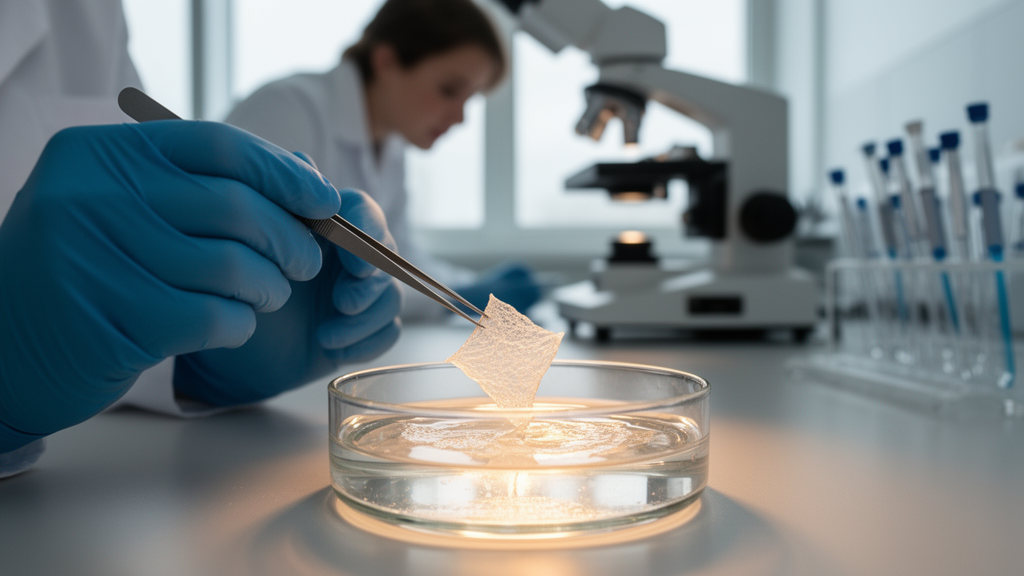
Deep wounds in mammals do not simply knit themselves back together the way some invertebrate tissues do. Instead, the body deploys specialized wound fibroblasts that arise from the fascia, the deepest connective tissue layer of the skin, and these cells drive inflammation, scar formation and the mechanical contraction that pulls wound edges together. Tracking what these precursors actually do inside living fascia, in real time, has long been a technical headache for laboratory researchers. A team publishing in BMC Methods now describes a deceptively simple way around the problem: peel the fascia free, float it in a culture dish, and watch it contract on its own.
The method, called the fascia explant, or FE, system, involves excising roughly three to four millimetre pieces of fascia from the back skin of mice or from human samples and culturing them free-floating in serum-supplemented DMEM media for up to seven days. Over that period, the soft, jelly-like translucent tissue progressively contracts to approximately fifty percent of its initial area, forming a dense, opaque structure reminiscent of the wound contraction observed in living animals. Because the explant retains fibroblast precursors in their native microenvironment, the authors argue that the system captures causative events of inflammation, scarring and contraction that artificial skin equivalents and standard in vitro models have struggled to reproduce.
The protocol is standardized for mice of any age, sex or genetic lineage, and it can likewise accommodate human donor tissue, which is collected and stored in cold DPBS before processing. Preparation itself is deliberately low-tech: the dorsal skin is shaved, treated with hair removal cream, excised with a sterile scalpel and gently peeled so that the superficial fascia stays attached to the skin rather than the underlying muscle. The translucent fascia layer is then lifted away, trimmed into explants, and transferred into wells of a six-well plate containing three millilitres of complete media, with three explants per well. Smaller formats work too, including 12-, 24- and 48-well plates, the latter suited to high-throughput screening with one explant per millilitre of media.
Culture conditions are equally conventional. Plates go into an incubator at 37 degrees Celsius, 21 percent oxygen, five percent carbon dioxide and 95 percent humidity. Day zero marks the start of the experiment, when plates are imaged under a bright-field microscope, a step repeated at equal intervals every day. Fresh pre-warmed media replaces the old on days two and four, along with any treatment compounds, and the authors caution that about 100 microlitres of media should remain in each well so tissues do not stick to the plastic or the pipette. The authors also recommend using a one-millilitre pipette rather than a vacuum pump for aspiration, because fascia explants are easily aspirated along with the spent media.
The readouts come in two flavors. Temporal information comes from the daily bright-field images, which document how a flat, translucent sheet of fascia rounds up into an opaque, scar-like disc. Spatial information comes at the end of the assay, when explants are fixed in two percent paraformaldehyde, dehydrated through sucrose gradients, embedded in OCT compound, frozen and cryo-sectioned at six to seven micrometres. Standard histology then reveals what has happened inside: Sirius red or Masson’s trichrome staining visualizes collagen, while immunofluorescence identifies fibroblast differentiation states within the explant.
Those differentiation dynamics follow a sequence consistent with in vivo wound healing. At day three of culture, fibroblasts within the explants predominantly show a pro-inflammatory phenotype, marked by expression of Podoplanin, or PDPN, while differentiation into the contractile state remains limited, with minimal expression of contractile markers. By day six, the majority of fibroblasts have transitioned into a contractile alpha-smooth muscle actin-positive myofibroblast state, accompanied by a reduction in the pro-inflammatory population. Because the timing of these stages is reproducible, researchers can simply limit the culture duration to capture intermediate steps of fibroblast differentiation that would otherwise be difficult to observe.
The system also responds to pharmacological manipulation in ways that mirror known wound biology. Transforming growth factor beta and Yes-associated protein signaling are among the most prominent pathways that activate fibroblasts and push them toward contractile states. Adding TGF-beta to the culture significantly accelerated contraction of the fascia explants, whereas blocking YAP activity with the inhibitor Verteporfin prevented tissue contraction altogether, compared with untreated controls. Quantification of explant area and contraction rates confirmed significant changes under these interventions, providing a proof of concept that fascia explants can identify modulators of wound healing, whether small molecules, recombinant proteins or neutralizing antibodies.
Several extensions broaden the platform’s reach. Fascia can be harvested from transgenic mouse lines, such as Engrailed-1 Cre mice crossed with fluorescent reporter lines, allowing direct observation of wound fibroblast precursors at single-cell resolution. Explants can be co-cultured with other cell types, including immune cells such as neutrophils and monocyte-macrophages, making the system suitable for studying immune-fibroblast interplay, a persistent shortcoming of existing in vitro assays. The authors also note that the translucent tissue is compatible with advanced three-dimensional imaging approaches such as confocal or light-sheet microscopy; while not applied to fascia explants alone in this study, their recent work embedding explants in matrigel demonstrated visualization of fibroblast protrusions and spatial organization in three dimensions.
Physiological context matters as well. Explants derived from young, aged and diabetic skin showed distinct contraction profiles, with tissue from young samples contracting rapidly and pronouncedly, while aged and diabetic samples displayed comparatively reduced or altered contractile responses. These differences emerged in ex vivo culture without any external modulation, which the authors interpret as evidence that the explants preserve intrinsic, context-dependent properties of their tissue of origin. Human fascia explants contracted similarly to murine ones over time, and the authors report that age and genetic strain, such as db/db mice, markedly influence contraction outcomes, so experimental design must account for these variables.
The authors are candid about the system’s limits. In this methods-focused study, they describe the phenotypic and functional features of the assay without directly assessing lineage preservation or identity, and they point out that the current culture conditions are normoxic at 21 percent oxygen, whereas the native sub-dermal microenvironment is relatively hypoxic; future studies under controlled low-oxygen conditions may reveal how oxygen tension modulates fibroblast activation and contraction. Technical care is also essential: residual hair or blood can interfere with contraction readouts, and accidentally excising fat or muscle along with the fascia can skew contraction rates. Still, the team positions the FE system as a versatile, scalable assay for drug candidate screening, agonist and antagonist testing, neutralizing antibody work and viral approaches, offering researchers a practical window into the cellular choreography of wound repair.
Subject of Research: A protocol for culturing fascia explants to study fibroblast differentiation and tissue contraction during wound healing
Article Title: Fascia explants enable real-time dynamic studies of wound fibroblast ancestry in living tissues
Article References: Dasgupta, B., Correa-Gallegos, D., Lee, A., & Rinkevich, Y. (2026). Fascia explants enable real-time dynamic studies of wound fibroblast ancestry in living tissues. BMC Methods, 3(1), Article 31. https://doi.org/10.1186/s44330-026-00084-5
Image Credits: AI Generated
DOI: 10.1186/s44330-026-00084-5
Keywords: fascia explants, wound healing, fibroblasts, tissue contraction, fibrosis, myofibroblasts, TGF-beta, YAP, drug screening, immunofluorescence, ex vivo culture, protocol
News Source: Gregory Coleman. (October 30, 2026). Floating Fascia Explants Let Researchers Watch Wound Fibroblasts Contract in Real Time. Scienmag.



