


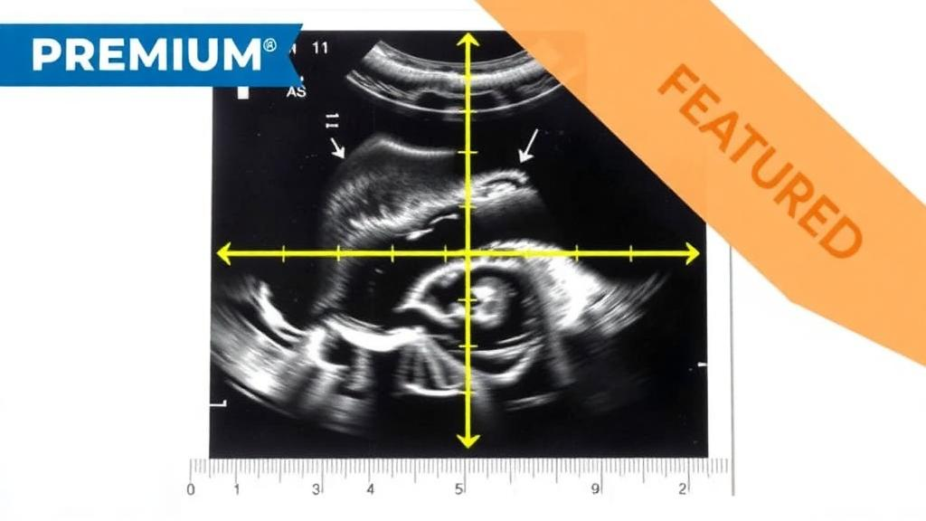
Quantitative ultrasound measurements of the cervix in pregnant women at risk for preterm birth can be repeated reliably by the same sonographer and reproduced across different sonographers, but only for some of the parameters tested, according to a prospective single-center study published in Discover Imaging. Two of six quantitative ultrasound (QUS) measurements — the attenuation coefficient and the Lizzi-Feleppa midband — reached moderate reliability benchmarks when averaged over four ultrasound acquisitions, while the remaining four parameters fell short and would need refinement before any clinical use.
The study, conducted at the University of Illinois Chicago between April 2018 and June 2022, addressed a gap that has limited the translation of QUS from the laboratory to prenatal care. QUS extracts quantitative information about tissue microstructure from the raw radiofrequency signals behind ordinary ultrasound images, rather than relying on the visual appearance of B-mode pictures. Previous work by the same group had suggested that QUS features of cervical tissue could help identify women at risk of spontaneous preterm birth, but earlier reliability studies had been performed on reference phantoms or excised tissue, not in living patients. Without evidence that the measurements are consistent when repeated in the body, no imaging biomarker can move toward routine clinical use.
Participants were drawn from a parent study of 529 pregnant women enrolled to test QUS technology for identifying women at risk for spontaneous preterm birth. Eligibility required a singleton pregnancy and the ability to read and speak English; women with multiple gestations, chronic medical conditions such as diabetes or hypertension, cervical cerclage, or major fetal anomaly were excluded. Each participant received standard clinical care plus research transvaginal scans at 20 plus-or-minus 2 and 24 plus-or-minus 2 weeks of gestation. In total, 81 participants with a mean age of 27.12 years were recruited, producing 82 examination pairs: 36 pairs in which the same sonographer performed both examinations, and 46 pairs performed by two different sonographers.
The scanning protocol was deliberately aligned with routine clinical practice. Registered diagnostic medical sonographers used a Siemens Acuson S2000 with MC9-4 transvaginal probes, placing the focus on the cervix at an imaging depth of 5 to 6 centimeters without pressing on the cervix and without any specific probe angle. Each research examination consisted of 10 consecutive transvaginal acquisitions plus one scan of a reference phantom used to calibrate the cervical data and adjust for system-dependent effects. Between the two examinations at a visit, participants dressed, walked down a corridor, and returned, so the repeat scan captured realistic re-examination conditions rather than a continuous back-to-back measurement.
Six QUS parameters were computed from the radiofrequency data within a field of interest drawn by the sonographer to capture homogeneous cervical tissue from the external to the internal os. The attenuation coefficient quantifies how much ultrasound energy the tissue absorbs and scatters, and was chosen because collagen remodeling and hydration changes during pregnancy alter these properties. The Lizzi-Feleppa intercept, midband, and slope derive from linear regression of decibel-scale backscatter coefficients against frequency and reflect the size and distribution of scatterers in cervical collagen. Envelope Kappa and Mu, parameters of a homodyned K distribution fitted to the signal envelope, relate to the ratio of coherent to incoherent scattering and to scatterer number density, features expected to change as cervical tissue softens.
The analysis began by testing whether transducers, phantoms, or research visits contributed significant variability. Using linear mixed models on first-acquisition data, the researchers found that transducer and reference phantom variances were not statistically significant, and the sonographer variance component was likewise not significant for any QUS measurement. Participant-to-participant differences and measurement error dominated the variance for all six parameters. This matters because it indicates the reference phantom calibration approach successfully removed instrument-related variation, leaving biological differences between women as the main source of spread in the measurements.
Repeatability and reproducibility were then quantified using the coefficient of variation, intraclass correlation coefficients, and Bland-Altman plots, with ICC benchmarks of above 0.9 for excellent, above 0.75 for good, above 0.5 for moderate, and below 0.5 for poor. When the first four acquisitions of each examination were averaged, attenuation coefficient, LF midband, LF slope, Kappa, and Mu showed moderate intra-sonographer repeatability, with coefficients of variation between 11.9 and 12.9 percent and ICCs between 0.62 and 0.69. LF intercept was the exception, with poor repeatability despite a low coefficient of variation of 6.9 percent, returning an ICC of only 0.38. The authors note that LF slope and LF intercept are inherently more variable than the midband because they come from the same linear regression derivation.
Across sonographers, the picture narrowed further. Attenuation coefficient and LF midband again achieved moderate inter-sonographer reproducibility, with coefficients of variation of 10.4 to 13.9 percent and ICCs of 0.61 to 0.63. LF intercept sat at the boundary with marginal reproducibility, an ICC of 0.51 and a coefficient of variation of 7.6 percent. Kappa and Mu performed poorly, with ICCs of 0.27 to 0.38 despite coefficients of variation of 6.9 to 14.5 percent, and LF slope also showed poor reproducibility with a moderately high coefficient of variation of 20.2 percent. Bland-Altman analysis confirmed these patterns, and assumption checks found non-normality only for a few parameter subsets, apparently driven by single outlier measurements, while a heteroscedasticity finding for LF midband did not survive adjustment for multiple testing.
The choice of four acquisitions was not arbitrary. The researchers computed ICCs for averages of the first k acquisitions, from one to ten, and found that averaging four acquisitions was sufficient to reach moderate ICC values for attenuation coefficient and LF midband, with single-acquisition results weaker across every measure. Because clinical guidelines already call for at least three cervical length measurements during a transvaginal examination, obtaining four acquisitions would be feasible in practice, making the averaging strategy realistic rather than burdensome.
The authors conclude that averaged in vivo transvaginal QUS measurements of attenuation coefficient and LF midband have potential for development as noninvasive clinical measurements with moderate reproducibility for auxiliary monitoring of pregnancy progress, while the other parameters require further refinement before they can be recommended. They frame this as a step toward using QUS as a complementary feature alongside cervical length in preterm birth risk models, noting that their earlier outcome studies analyzed only one of ten acquisitions per visit, so repeated acquisitions could improve prediction. The study has clear limitations: it was single-center, recruitment into the repeatability portion was non-systematic because of COVID-19 restrictions and scheduling constraints, no formal record of women approached or declined was kept, and the sample did not support meaningful subgroup comparisons across racial and ethnic groups, even though participants were diverse. All research activity was suspended from March 2020 to February 2021, and for the following year only one sonographer could scan at a time, which constrained the mix of same- and cross-sonographer pairs. This was a measurement reliability study in 81 women at 20 to 24 weeks of gestation; it did not test whether QUS measurements actually predict preterm birth outcomes, and the authors themselves state that future studies are needed to determine whether training and protocol standardization can improve the reliability values further.
Subject of Research: Repeatability and reproducibility of transvaginal quantitative ultrasound cervical tissue measurements in pregnant women at risk for preterm birth
Article Title: Repeatability and reproducibility of quantitative ultrasound cervical measurements in women at risk for preterm birth
Article References: Villegas-Downs, M., Gao, Z., Jia, H., Han, A., McFarlin, B. L., O’Brien, W. D., Jr., & Simpson, D. G. (2025). Repeatability and reproducibility of quantitative ultrasound cervical measurements in women at risk for preterm birth. Discover Imaging, 2(1), Article 16. https://doi.org/10.1007/s44352-025-00020-3
Image Credits: AI Generated
DOI: 10.1007/s44352-025-00020-3
Keywords: quantitative ultrasound, preterm birth, cervix, pregnancy, repeatability, reproducibility, intraclass correlation, transvaginal ultrasound, attenuation coefficient, biomarkers, prenatal screening, Bland-Altman analysis
News Source: Harold Sullivan. (October 29, 2026). Quantitative Ultrasound Cervix Measurements Show Moderate Reliability in Pregnancy Study. Scienmag.



