


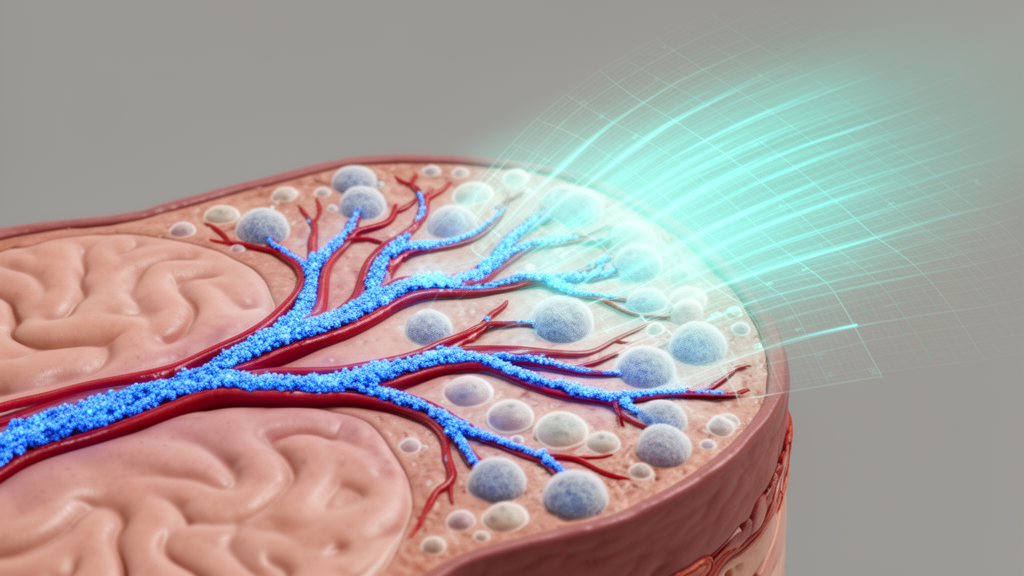
A new study published in Acta Neuropathologica Communications reports a digital pathology approach that automates the detection of cortical CAA-positive vessels separately from β-amyloid plaques, validates the method against expert manual scoring, and maps how closely plaques sit to affected vessels. The work was conducted on digitized histopathologic slides from the calcarine cortex, a region of the visual cortex at the back of the brain.
The research team, led by Paniz Mojdeganlou and senior author Alifiya Kapasi, trained machine-learning classifiers on the HALO AI platform to tell CAA-positive vessels apart from β-amyloid plaques on the digitized slides. They applied the classifiers to 100 slides that expert neuropathologists had previously rated by eye. From the digital output the team derived quantitative measures for CAA — the CAA classified area and the vascular β-amyloid percent positivity — and for plaques, measured as β-amyloid plaque classified area. These digital values were then compared with the manual CAA scores and with ratings of Alzheimer’s disease neuropathologic change, or ADNC.
The validation results were strong. Digital CAA classified area correlated with semi-quantitative manual CAA scores at a Spearman coefficient of 0.96, and vascular β-amyloid percent positivity correlated at 0.79. The counts of CAA-positive vessels per slide ranged from 0 to 820, with a median of 3 vessels in manual score 1, 12 in score 2, 153 in score 3, and 399 in score 4 — a steep gradient showing that the digital counts tracked increasing severity. The participants whose tissue was studied had a mean age of 91.1 years at death, with a standard deviation of 5.6, and 76% were women.
The plaque-side measurements also performed well, though somewhat less strikingly. Digital quantification of β-amyloid plaque burden correlated strongly with Thal phase, a standard staging measure of amyloid spread, at r = 0.80, and moderately with ADNC at r = 0.60. Together these coefficients indicate that a single automated pass over a slide can capture both the vascular and the plaque components of amyloid pathology in a way that mirrors expert consensus ratings.
The most intriguing finding came from the spatial analysis. The total β-amyloid plaque area was relatively similar across increasing manual CAA scores — in other words, cases with severe CAA did not simply have more plaque overall. But when the researchers defined perivascular plaques as β-amyloid plaques lying within a concentric distance of 0 to 150 microns from the center of a CAA-positive vessel, a clear pattern emerged: cases with higher CAA scores had more perivascular plaques closest to CAA-positive vessels. This suggests that the local relationship between vessels and plaques, rather than total plaque burden, is what changes with CAA severity.
That spatial proximity measure is potentially significant for understanding how CAA develops. The study’s stated conclusion is that the digital classifiers reliably distinguish cortical CAA vessels from β-amyloid plaques and can be utilized to study plaque proximity to CAA-positive vessel severity, supporting digital pathology as a means of advancing understanding of CAA pathogenesis.
The study’s scope and limitations are worth keeping in view. The classifiers were trained and validated on slides from a single cortical region, the calcarine cortex, in a cohort of very old donors, so performance in other brain regions or younger populations remains to be established. The correlations with manual scores describe agreement with existing semi-quantitative ratings, not diagnostic accuracy against an independent gold standard. And because the analysis is cross-sectional autopsy tissue, the proximity findings show an association between CAA severity and plaque location, not a causal sequence over time.
Even with those caveats, the methodological advance is concrete. Replacing eyeball estimates of vessel amyloid with reproducible digital measurements — down to counting individual CAA-positive vessels and measuring plaque distance from vessel centers in microns — gives researchers a far finer-grained instrument than the four-point manual scales in common use. The authors note the article is being shared early as a peer-reviewed, accepted, citable version with a permanent DOI, subject to further edits before the final Version of Record.
Subject of Research: Digital pathology quantification of vascular β-amyloid and perivascular plaques in cerebral amyloid angiopathy
Article Title: Digital quantification of vascular β-amyloid and perivascular plaques in cerebral amyloid angiopathy
Article References: Mojdeganlou, P., Poirier, J., Barnes, L. L., Bennett, D. A., Leurgans, S. E., Schneider, J. A., & Kapasi, A. (2026). Digital quantification of vascular β-amyloid and perivascular plaques in cerebral amyloid angiopathy. Acta Neuropathologica Communications. https://doi.org/10.1186/s40478-026-02450-4
Image Credits: AI Generated
DOI: 10.1186/s40478-026-02450-4
Keywords: cerebral amyloid angiopathy, β-amyloid, digital pathology, HALO AI, perivascular plaques, neuropathology, Alzheimer's disease, Thal phase, machine learning, calcarine cortex, autopsy tissue, validation
News Source: Diana Fleming. (October 29, 2026). Digital Pathology Tool Quantifies Amyloid Vessels and Plaques in CAA. Scienmag.



