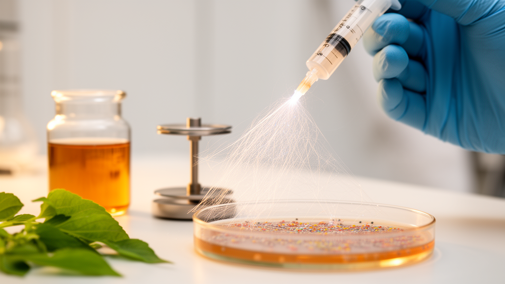
Wound dressings that can fight infection while keeping the wound environment moist have long been a goal of biomaterials research, and a new study published in the journal Fashion and Textiles on 30 June 2026 reports a laboratory-scale route to one such candidate material. Researchers Yujin Lee and Jungsoon Lee fabricated nanofibers from poly(vinyl alcohol), or PVA, loaded with 20 wt% Sophorae fructus extract, a preparation derived from the fruits of Sophora japonica L., a tree widely distributed across East Asia. The extract is rich in flavonoids and isoflavonoids such as genistein, rutin, sophoricoside, and kaempferol derivatives, compounds that have been reported to show antioxidant, antibacterial, anti-inflammatory, antiviral, and hemostatic activities. By electrospinning the polymer-extract solution into fibers with diameters in the nanometer range and then applying carefully controlled heat treatment, the team produced flexible fiber mats whose properties they systematically measured, identifying 170 °C for 45 minutes as the optimal cross-linking condition under their experimental settings.
The choice of PVA as the carrier polymer reflects both its strengths and its weaknesses. PVA is hydrophilic, biocompatible, and non-toxic, and its excellent film-forming ability and mechanical flexibility make it well suited to electrospinning, a technique that applies high voltage to a polymer solution to draw out fibers far thinner than those achievable by conventional weaving. The high specific surface area and network structure of electrospun nanofibers make them skin-friendly and therefore attractive for wound dressing applications. The problem is water: PVA dissolves readily in moist environments, which is exactly the condition at a wound site. Chemical cross-linking agents such as glutaraldehyde can improve water resistance but raise potential toxicity concerns. Heat treatment offers a simpler, chemical-free alternative, promoting physical cross-linking by strengthening intermolecular hydrogen bonding and increasing polymer crystallinity. Too much heat, however, can fuse fibers together and destroy the porous architecture that makes nanofiber mats useful.
To make the material, the researchers extracted dried Sophorae fructus with water at a solid-to-liquid ratio of 1:20 at 100 °C for 60 minutes, repeating the extraction three times and concentrating the combined filtrates into a gum-like extract. They dissolved PVA in water at 11 wt%, added SFE at 20 wt% relative to the polymer content, stirred the mixture for more than 12 hours, and sonicated it for one hour to ensure homogeneity. Electrospinning was carried out at an applied voltage of 15 kilovolts, a tip-to-collector distance of 15 centimeters, and a flow rate of 2 milliliters per hour.
Microscopic examination with field-emission scanning electron microscopy revealed how the fiber architecture evolved with heat input. Untreated fibers displayed a uniform structure with smooth surfaces and clear boundaries between fibers. As temperature and duration increased, wave-like deformation and partial fusion at fiber contact points became progressively more pronounced, and the average fiber diameter grew accordingly. Interestingly, the two variables did not contribute equally: fibers treated at 160 °C for 60 minutes showed greater thermal deformation than those treated at 170 °C for 45 minutes, showing that prolonged exposure matters, while fibers treated at 180 °C for 30 minutes deformed more severely than the 170 °C intermediate, showing that sufficiently high temperature dominates. At 180 °C, localized melting even produced partial film-like structures. Only the fibers treated at 170 °C for 45 minutes retained their fibrous network and flexibility, qualities the authors describe as essential for wound dressing use.
Mechanical testing on a universal testing machine followed ASTM standards with Type IV specimens and a 25 millimeter gauge length, each sample tested in triplicate. The initial Young’s modulus and tensile strength generally rose with increasing heat-treatment severity, reflecting the enhanced intermolecular interactions and crystallinity induced by thermal cross-linking. Tensile strength, however, peaked at the 170 °C for 45 minute condition and then declined under harsher treatments such as 180 °C for 30 minutes or longer, because rapid crystallization and partial fusion at that temperature leave little further benefit from extended exposure. Elongation at break decreased progressively with heat-treatment severity, a consequence of moisture removal, the replacement of water-mediated hydrogen bonds with direct bonds between PVA chains, and the resulting loss of chain mobility. The 170 °C for 45 minute treatment delivered the most balanced mechanical profile, combining relatively high tensile strength and elongation with a moderate modulus, achieving sufficient cross-linking without excessive embrittlement.
Porosity, which governs cell infiltration, nutrient diffusion, oxygen permeability, and exudate management in a dressing, told a similar story of trade-offs. Measured by liquid displacement with ethanol, porosity fell from approximately 83% in untreated fibers and 79% after treatment at 160 °C for 30 minutes, a difference that was not statistically significant, to roughly 59% at 160 °C for 60 minutes, 51% at 170 °C for 45 minutes, and only about 36% and 31% at 180 °C for 30 and 60 minutes respectively. Because the optimal porosity range for wound healing is generally reported as 40 to 90%, the 180 °C samples fell outside the desirable range even at the shorter treatment time. Temperature proved more damaging to porosity than time, likely because fibers soften and partially melt quickly at higher temperatures. Water vapor transmission rate, by contrast, was largely unaffected: most heat-treated samples showed slightly increased WVTR values that reached the range of 2000 to 2500 grams per square meter per day considered optimal for wound healing, with no statistically significant differences among most heat-treated samples despite the porosity losses.
Water retention and degradability tests in phosphate-buffered saline at 37 °C exposed the consequences of inadequate cross-linking most dramatically. Untreated fibers and those treated at 160 °C dissolved immediately in the buffer, demonstrating insufficient aqueous stability for any practical dressing use. In contrast, fibers treated at 170 °C for 45 minutes, 180 °C for 30 minutes, and 180 °C for 60 minutes remained intact, with swelling ratios of approximately 823%, 538%, and 379% respectively, water retention declining as treatment severity increased. Degradation behavior followed a parallel pattern. Since an ideal dressing should remain intact for at least 14 days while tissue regeneration and angiogenesis proceed, the degradation timeline matters: fibers treated at 170 °C for 45 minutes degraded completely by the second week, while those treated at 180 °C for 30 and 60 minutes degraded far more slowly, retaining substantial residual mass after three weeks. Scanning electron microscopy of partially degraded samples showed extensive fiber fusion and film-like morphology in the 180 °C for 30 minute specimens, while the 180 °C for 60 minute samples retained more distinct fibrous features.
The biological performance of the mats revealed both a cost and a resilience of heat treatment. In the ABTS radical scavenging assay, heat-treated samples showed reduced antioxidant activity compared with untreated fibers, with greater reductions at higher temperatures and longer durations. Fibers treated at 160 °C for 30 and 60 minutes and at 170 °C for 45 minutes approached nearly 100% radical scavenging after 150 minutes, whereas those treated at 180 °C reached only about 94% and 81% for 30 and 60 minutes respectively. The authors attribute this decline to increased crystallinity and reduced diffusion pathways within the fiber matrix, which hinder the release of antioxidant components from the extract. Antibacterial activity against Staphylococcus aureus, one of the most prevalent bacterial species in chronic wounds, told a different story. Evaluated by the plate count method following ISO 20743:2021, all samples containing 20 wt% SFE showed antibacterial activity regardless of heat-treatment conditions, with no significant differences before and after treatment, indicating that the antibacterial constituents of the extract remained thermally stable under the applied conditions.
Taken together, the results map a clear trade-off between structural stability and functional performance, and they point to 170 °C for 45 minutes as the condition that best balances the competing requirements. This treatment provided water resistance, strong mechanical properties, controlled degradability over roughly two weeks, porosity within the desirable range, preserved moisture permeability, high antioxidant release, and stable antimicrobial performance, all while largely preserving the fibrous morphology that gives the mats their flexibility. The authors emphasize that the optimal condition is determined not by any single property but by the balance between mechanical reinforcement and functional behavior, and they suggest that heat-treated SFE/PVA nanofibers are promising candidates for advanced wound dressings, including potential use in demanding environments where material reliability, safety, and biocompatibility are critical. As with all laboratory studies, these findings come from in vitro characterization rather than clinical testing, so the materials remain candidates whose real-world wound healing performance would need to be established through further evaluation.
Subject of Research: Heat-treated electrospun PVA nanofibers loaded with Sophora fructus extract for antibacterial wound dressings
Article Title: Fabrication of Sophora fructus loaded polyvinyl alcohol nanofibers for antibacterial wound dressings
Article References: Lee, Y., & Lee, J. (2026). Fabrication of Sophora fructus loaded polyvinyl alcohol nanofibers for antibacterial wound dressings. Fashion and Textiles, 13(1), Article 28. https://doi.org/10.1186/s40691-026-00473-y
Image Credits: AI Generated
DOI: 10.1186/s40691-026-00473-y
Keywords: electrospinning, nanofibers, polyvinyl alcohol, Sophora fructus, wound dressings, antibacterial, heat treatment, cross-linking, porosity, antioxidant, Staphylococcus aureus, biomaterials
News Source: Denise Maddox. (October 29, 2026). Plant Extract Meets Electrospinning: Heat-Tuned Nanofibers Emerge as Antibacterial Wound Dressing Candidates. Scienmag.