


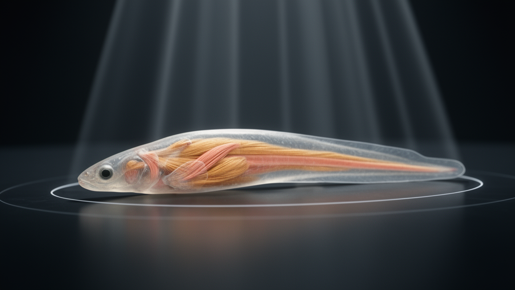
Using a battery of advanced three-dimensional imaging techniques, researchers have produced the most detailed reconstruction yet of the cranial muscles in lamprey larvae, uncovering a previously undescribed upper lip muscle and revealing that most lower lip muscles receive nerve supply from more than one branch of the trigeminal nerve.
The origin of the vertebrate jaw remains one of the most enduring puzzles in evolutionary biology. Because lamprey larvae retain fundamental anatomical and developmental features shared with hagfish that are considered ancestral for cyclostomes, they have been central to debates over how the jawless vertebrate head was transformed into the jawed condition. Yet their cranial anatomy has remained contentious, largely because earlier studies relied on gross dissection and histological sectioning, methods that left considerable room for subjective identification and produced descriptions that were inconsistent with one another.
To resolve these uncertainties, a team led by Ken-ichiro Yoshimoto and Daichi G. Suzuki examined large ammocoete larvae of the Northern Japanese brook lamprey, Lethenteron mitsukurii, collected from the Kamo River in Toyama, Japan, together with sea lamprey larvae, Petromyzon marinus, from a tributary of the Miño river in Spain.
Each method contributed a distinct strength. The contrast-enhanced micro-CT, scanned at a resolution of 7.7 micrometers, proved best for resolving fine details such as the directions of muscle fibers. Phase-contrast X-ray imaging, with a voxel size of 6.5 micrometers, and synchrotron micro-CT, at 2.26 micrometers the finest resolution in the study, visualized soft tissues without any contrast agents, avoiding the shrinkage that staining can cause. Light-sheet microscopy of tissue cleared with the CUBIC protocol and stained for acetylated tubulin allowed the trigeminal nerve branches to be reconstructed in three dimensions, something none of the CT methods could achieve. The researchers then manually segmented muscles and nerves in the software Amira, merging the datasets so that muscles and their innervation could be viewed simultaneously.
The reconstructions identified thirteen mandibular muscles and one hyoid muscle in L. mitsukurii. The same number of mandibular muscles was found in the sea lamprey, with no obvious interspecific differences, leading the authors to suggest that the arrangement they documented likely represents the ancestral condition of crown lamprey larvae. Among the upper lip muscles was a previously unreported one, named m. elevator labii superioris, a very thin muscle beneath the larger m. buccalis superficialis that is clearly visible in sagittal images but easy to miss in transverse or coronal sections.
The innervation findings are among the most consequential. The branch termed rV2/3A suborbitalis had long been thought to be purely sensory, but the new data show it supplies the newly identified upper lip muscle, indicating it contains motor components. The rV2/3A suprapharyngeus branch, whose motor role had also been debated, was confirmed to innervate the muscle m. retractor labialis dorsalis. In the lower lip, the picture is even more complex: the m. retractor papillaris and m. retractor labialis ventralis are each innervated by two trigeminal subbranches, rV2/3A apicalis II and rV2/3B, while the m. elevator labialis ventralis receives a triple innervation from rV2/3A apicalis II, rV2/3A subpharyngeus and rV2/3B, a pattern the team confirmed with immunostained histological sections.
This dual and triple innervation matters for understanding how the vertebrate jaw evolved. In jawed vertebrates, motor components of the trigeminal nerve are restricted to the mandibular branch, which controls the jaw-closing muscles. In lampreys, motor fibers run in both rV2/3A and rV2/3B, and the new findings show that the motor innervation of the lower lip cannot be assigned unambiguously to either branch. The authors interpret the lower lip as the interface between two developmental modules of the mandibular arch, an anterior oral module associated with rV2/3A and a posterior velar module associated with rV2/3B, with the overlapping nerve supply reflecting this interfacial position. Multiple motor innervation of muscles, they note, is not uncommon at boundaries between developmental modules, as seen in the cucullaris muscles of jawed vertebrates and the intermandibularis of some elasmobranchs and lungfishes.
The detailed topography of the lower lip also yielded an unexpected evolutionary clue. The m. elevator labialis ventralis originates from the ventromedial longitudinal bar, the very site where the piston cartilage of the adult lingual apparatus develops during metamorphosis. Its ventral portions run anteroposteriorly, matching the orientation of adult lingual muscles, and its innervation pattern resembles that of adult dorsal lingual muscles, one of which is doubly innervated in a comparable way. Taken together, these affinities suggest that this larval lower lip muscle is a precursor of the adult lingual musculature, the piston-like feeding apparatus of parasitic adult lampreys that is not homologous to the tongue of jawed vertebrates. Earlier studies had overlooked this connection because the muscle was misidentified and illustrated only in lateral view.
The velar muscles were likewise redrawn. The velum, a pumping structure that generates respiratory water flow in the larva and degenerates to a simple anti-reflux valve in adults, is derived from the mandibular arch and supplied by rV2/3B. The team distinguished five velar muscles and showed that Tretjakoff and Damas had mistakenly split or merged several of them, for example describing the ventral portion of m. velaris dorso-ventralis as a separate muscle. Because well-preserved fossils of jawless stem gnathostomes such as galeaspids and osteostracans show a large mandibular fossa that some paleontologists have interpreted as housing a pumping velum, the new quantitative three-dimensional models of the larval velum offer a benchmark for testing whether such a structure existed in the ancestors of jawed vertebrates, or whether the velum is instead a cyclostome novelty.
The authors caution that each method has limitations. Iodine staining can shrink tissue, though comparable morphology from unstained scans suggests the effect was minor here. Light-sheet imaging required skin removal and chemical clearing, procedures that risk deforming superficial muscles and nerves, and light scattering blurred deeper regions, so cross-referencing with CT images and histological sections was essential. The team also found no trace of a nerve branch, the r. basilaris, that earlier workers reported supplying the superficial buccal muscle, and they note that interspecific variation, detection limits or preparation loss remain possible explanations. Because the specimens were fixed larvae, the study documents anatomy rather than function, and the probable functions assigned to each muscle are inferences from their positions. The researchers argue that combining these non-destructive imaging methods with gene expression analysis on the same samples offers a promising route for tracking how larval cranial muscles are remodeled into the adult head during metamorphosis, a process that remains poorly described and that bears directly on how the jawed vertebrate skull came to be.
Subject of Research: Three-dimensional imaging-based redescription of cranial muscle anatomy and trigeminal innervation in lamprey larvae
Article Title: Three-dimensional detailed redescription of the lamprey larval cranial muscles based on multiple imaging methods
Article References: Yoshimoto, K.-I., Kariyayama, H., Tamura, M., Koyabu, D., Pérez-Fernández, J., Yoneyama, A., Hyodo, K., Hirano, K., Nonaka, S., Higashiyama, H., Hirasawa, T., & Suzuki, D. G. (2026). Three-dimensional detailed redescription of the lamprey larval cranial muscles based on multiple imaging methods. Developmental Biology Advances, 17(1), Article 18. https://doi.org/10.1186/s13227-026-00271-z
Image Credits: AI Generated
DOI: 10.1186/s13227-026-00271-z
Keywords: lamprey larvae, cranial muscles, trigeminal nerve, micro-computed tomography, light-sheet microscopy, vertebrate jaw evolution, ammocoetes, velum, lingual apparatus, metamorphosis, cyclostomes, comparative morphology
News Source: Gavin Prescott. (October 29, 2026). Hidden Cranial Muscles of Lamprey Larvae Revealed in 3D Imaging Study. Scienmag.



