A previously published electron microscopy dataset has yielded new findings in a second life. Researchers reanalyzed publicly available high-resolution transmission electron microscopy images from an earlier umbelliferone study and found that the compound was associated with changes in the postsynaptic density that the original publication had not described.
The original study examined umbelliferone (UMB) under scopolamine (SCO)-induced pathological conditions and reported recovery of synaptic vesicle architecture. Its high-resolution electron microscopy dataset was deposited in the Korea BioData Station (K-BDS), a national biological data platform, making a secondary analysis possible without new experiments.
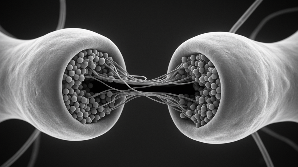
In the new study, published in Applied Microscopy, the team turned to a structure the original work had not explored: the postsynaptic density (PSD), a structural determinant of synaptic efficacy and plasticity. Using the original images, they performed quantitative secondary analyses of PSD length, width, and ultrastructural features across the experimental groups.
The reanalysis showed that UMB treatment significantly increased PSD length and width compared with the SCO-treated group, which the authors interpret as indicating structural restoration of impaired postsynaptic architecture. These PSD-level alterations had not been described in the original publication.
The authors emphasize that the finding illustrates how purposeful reuse of existing data can generate novel biological insights. Public repositories, they note, not only support reproducibility and transparency but also enable discoveries beyond the scope of the original studies.
The work also serves as a demonstration of K-BDS itself. By showing that a deposited dataset could support a new line of analysis, the study underscores the platform’s role in facilitating secondary discovery and data-driven neuroscience research without additional animal experimentation.
Because the analysis is a reanalysis of existing microscopy images rather than a new experiment, its conclusions rest on the same imaging material and experimental design as the original study. The reported PSD changes are structural measurements from electron micrographs, not direct functional readouts of synaptic transmission.
Nevertheless, the study offers a concrete example of data reuse in neuroscience: a dataset deposited to satisfy open-science practices became the basis for a previously unreported observation about postsynaptic structure, obtained without any new animal work.
Subject of Research: Reanalysis of public electron microscopy data showing umbelliferone-associated changes in postsynaptic density structure
Article Title: Umbelliferone-induced recovery of postsynaptic density structure revealed by the reanalysis of public electron microscopy data
Article References: Choi, G.-Y., Choi, H., Lee, S., & Kweon, H.-S. (2026). Umbelliferone-induced recovery of postsynaptic density structure revealed by the reanalysis of public electron microscopy data. Applied Microscopy, 56(1), Article 17. https://doi.org/10.1186/s42649-026-00129-7
Image Credits: AI Generated
DOI: 10.1186/s42649-026-00129-7
Keywords: umbelliferone, scopolamine, postsynaptic density, electron microscopy, data reuse, synapse, Korea BioData Station, neuroscience, open data, secondary analysis, Umbelliferone-induced, recovery
News Source: Denise Maddox. (October 29, 2026). Reanalysis of Public Electron Microscopy Data Reveals Umbelliferone’s Effects on Synaptic Structure. Scienmag.