


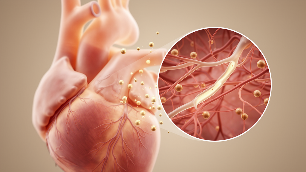
Heart failure with preserved ejection fraction, a condition in which the heart muscle stiffens and fills poorly even though the pumping fraction appears normal, may in some patients be best understood as a cardiac manifestation of diabetic microvascular disease. The authors emphasize that their proposal is a conceptual framework built on converging lines of evidence rather than a demonstration of a single causal pathway, and they argue that coronary microvascular dysfunction could serve as the mechanistic bridge between systemic metabolic stress and myocardial stiffening.
The scale of the problem is considerable. Heart failure with preserved ejection fraction, commonly abbreviated HFpEF, accounts for roughly half of all heart failure cases, and diabetes is highly prevalent within this population, affecting approximately 40 to 45 percent of patients with HFpEF according to the review. Epidemiological studies cited by the authors indicate that type 2 diabetes confers an approximately two- to five-fold increased risk of heart failure compared with non-diabetic populations. In the nationwide DIABET-IC cohort, heart failure was present in 39.2 percent of type 2 diabetes patients at baseline, and among those who developed heart failure during follow-up, 46.6 percent of newly diagnosed cases were of the preserved ejection fraction type, making it the predominant incident phenotype in that population. The incidence of heart failure reached 3.0 per 100 person-years in that cohort.
Preclinical disease appears widespread as well. Meta-analytic data reviewed by the authors show that among adults with type 2 diabetes, the pooled prevalence of HFpEF was 17 percent, while asymptomatic left ventricular diastolic dysfunction affected 43 percent of patients, suggesting a large reservoir of individuals who may progress to overt heart failure. Similar patterns extend to type 1 diabetes. In the Thousand & 1 Study, grossly abnormal systolic or diastolic function was identified in 15 percent of asymptomatic type 1 diabetes patients without known heart disease, and diabetic cardiomyopathy manifesting initially as diastolic dysfunction was reported in 34 percent of adult type 1 diabetes populations. The authors also caution that HFpEF is not a benign diagnosis: landmark population-based studies found that adjusted one-year mortality did not significantly differ between HFpEF and reduced-ejection-fraction populations, with a hazard ratio of 1.13 and a 95 percent confidence interval of 0.94 to 1.36, and that survival in HFpEF has remained largely unchanged over time even as outcomes improved for reduced ejection fraction.
The review’s core hypothesis rests on a striking overlap between the defining features of the two conditions. Classical diabetic complications, including nephropathy, retinopathy and neuropathy, are fundamentally microvascular in nature, characterized by endothelial dysfunction, capillary rarefaction, impaired nitric oxide signaling, oxidative stress and barrier disruption. In human kidney biopsy studies, glomerular basement membrane thickening, mesangial expansion and altered capillary architecture have been documented even at early clinical stages, and structural changes can precede overt proteinuria. Contrast-enhanced ultrasound studies have demonstrated delayed and reduced renal cortical perfusion in diabetic patients, with perfusion indices correlating with glomerular filtration rate. In the retina, pericyte loss, capillary microaneurysm formation and capillary occlusion define early disease, while in diabetic neuropathy, reduced perfusion of the vasa nervorum contributes to nerve ischemia. Across these organs, the earliest detectable abnormalities arise in capillary networks, with endothelial dysfunction as the common denominator.
Human studies show that this endothelial phenotype emerges early in diabetes. Impaired endothelium-dependent vasodilation, measured by flow-mediated dilation, has been reported even in normoglycemic individuals at risk for type 2 diabetes, and meta-analyses confirm significantly reduced flow-mediated dilation in established type 2 diabetes. Circulating adhesion molecules, including vascular cell adhesion molecule-1, intercellular adhesion molecule-1 and selectins, are elevated in type 2 diabetes and correlate with both prevalent and incident microvascular complications; in prospective analyses, baseline vascular cell adhesion molecule-1 independently predicted the later development of diabetic neuropathy, retinopathy and nephropathy. Endothelial insulin resistance, in which the vascular actions of insulin on vasodilation and microvascular recruitment are blunted, is detectable in obesity and type 2 diabetes before structural complications develop, and a 2025 study reported that obesity-associated elevations in adrenomedullin impair endothelial insulin receptor signaling and nitric oxide synthase activation. The endothelial glycocalyx, a regulator of vascular permeability, is significantly thinned in patients with diabetes, and its degradation correlates with albuminuria and other markers of microvascular damage.
Strikingly similar microvascular abnormalities are now recognized in HFpEF itself. In the prospective multinational PROMIS-HFpEF study, 75 percent of rigorously characterized HFpEF patients exhibited impaired coronary flow reserve, defined as a value below 2.5, even in the absence of unrevascularized epicardial coronary artery disease, and lower coronary flow reserve independently correlated with markers of systemic endothelial dysfunction, elevated NT-proBNP, right ventricular dysfunction and impaired myocardial strain. Invasive assessment reinforces the burden: in one study of hospitalized HFpEF patients, 85 percent had either epicardial coronary artery disease or coronary microvascular dysfunction, and 81 percent of those without obstructive coronary disease exhibited microvascular dysfunction. The abnormality carries prognostic weight, as cardiac magnetic resonance-derived coronary flow reserve below 2.0 independently predicted death or heart failure hospitalization and outperformed conventional structural markers. Autopsy-based work by Mohammed and colleagues demonstrated reduced microvascular density and increased interstitial fibrosis in HFpEF hearts, with fibrosis correlating inversely with microvascular density, while other studies identified coronary microvascular endothelial activation with increased adhesion molecule expression and NADPH oxidase-mediated oxidative stress.
A mechanistic framework articulated by Paulus and Tschöpe connects these microvascular findings to myocardial stiffness. In this model, cardiometabolic comorbidities induce coronary microvascular endothelial inflammation, reducing nitric oxide bioavailability, which lowers myocardial cGMP and protein kinase G activity, promoting titin hypophosphorylation, cardiomyocyte hypertrophy and increased passive stiffness. Human myocardial biopsy studies support the axis, showing significantly reduced myocardial protein kinase G activity in HFpEF associated with increased cardiomyocyte resting tension, and experimental models indicate that titin hypophosphorylation accounts for a major component of diastolic stiffness in metabolically driven HFpEF. Importantly, the review argues that the microvascular involvement extends beyond the heart. In HFpEF, skeletal muscle shows a shift from oxidative type I fibers toward glycolytic type II fibers, a reduced capillary-to-fiber ratio, impaired perfusion reserve, attenuated oxygen extraction and reduced mitochondrial oxidative capacity, all of which correlate with reduced exercise capacity. Albuminuria, a marker of glomerular endothelial injury, is independently associated with adverse outcomes in HFpEF, and in a contemporary cohort of 332 HFpEF patients, diabetes and albuminuria each independently predicted adverse outcomes, with their coexistence conferring an approximately threefold higher risk.
These converging mechanisms have therapeutic implications, and the review surveys how contemporary HFpEF therapies may intersect with microvascular biology. Sodium-glucose cotransporter 2 inhibitors reduce the composite of cardiovascular death or heart failure hospitalization across randomized trials, and in patients with type 2 diabetes and HFpEF they improved endothelial function measured by flow-mediated dilation, reduced arterial stiffness and improved diastolic indices, with improvements in diastolic function independently associated with changes in endothelial function and insulin resistance. In patients with type 2 diabetes and stable coronary artery disease, dapagliflozin significantly improved myocardial flow reserve over a short treatment period. In the STEP-HFpEF trial, once-weekly semaglutide significantly improved heart failure-related symptoms, six-minute walk distance and C-reactive protein alongside substantial weight reduction, with similar results in patients with concomitant type 2 diabetes in the STEP-HFpEF DM trial. The nonsteroidal mineralocorticoid receptor antagonist finerenone significantly reduced the composite of worsening heart failure events and cardiovascular death in the FINEARTS-HF trial, and in the mechanistic phase 2 IMPROVE-DiCE study, a metabolic intervention improved the myocardial phosphocreatine-to-ATP ratio in patients with obesity and type 2 diabetes, with or without HFpEF, although that study was limited by small sample size and open-label design.
Perhaps the most provocative recent evidence comes from experimental models in which endothelial-specific molecular perturbations can induce or ameliorate HFpEF phenotypes in diabetic settings. Endothelial overexpression of Rnd3 attenuated diabetic myocardial microvascular injury and improved cardiac dysfunction in streptozotocin and high-fat diet-induced diabetic HFpEF mouse models through Trim40-mediated ubiquitination and suppression of ROCK1 signaling. Endothelial SIRT6 deficiency enhanced fatty acid transport across the endothelial barrier and promoted myocardial lipid accumulation, while endothelial restoration of SIRT6 improved diastolic function in the same diabetic models, and endothelium-specific deletion of USP7 mitigated fibrosis in a pressure and metabolic stress model. Disruption of mitochondrial hexokinase-1 localization in endothelial cells drove O-GlcNAc-dependent metabolic remodeling associated with HFpEF-like phenotypes. Multi-omic and spatial transcriptomic analyses of the type 2 diabetic heart further reveal coordinated transcriptional reprogramming within coronary microvascular-enriched regions, including endothelial depletion, fibroblast expansion and fatty acid metabolic reprogramming arranged in structured regional patterns, although the authors note these datasets remain largely descriptive and causality is unresolved.
The authors conclude that cardiometabolic HFpEF, particularly in patients with diabetes and obesity, may share fundamental pathophysiological features with diabetic microvascular complications, and that strategies aimed at restoring endothelial homeostasis and microvascular integrity may represent a more mechanistically aligned therapeutic approach than targeting cardiomyocyte mechanics alone. They acknowledge that diabetes has traditionally been regarded primarily as a comorbidity in HFpEF rather than a driver of disease development, and that reframing HFpEF within the context of diabetic microvascular injury would require further prospective validation. If the framework holds, coronary flow reserve assessment and circulating endothelial biomarkers such as syndecan-1 and adhesion molecules, which remain incompletely standardized, could eventually improve phenotyping and therapeutic stratification in this large and growing patient population.
Subject of Research: The proposed role of microvascular dysfunction in linking diabetes to heart failure with preserved ejection fraction
Article Title: HFpEF as a diabetic complication: the central role of microvascular dysfunction
Article References: Nagai, Y., Matoba, K., & Nishimura, R. (2026). HFpEF as a diabetic complication: the central role of microvascular dysfunction. Discover Endocrinology and Metabolism, 2(1), Article 10. https://doi.org/10.1007/s44417-026-00023-6
Image Credits: AI Generated
DOI: 10.1007/s44417-026-00023-6
Keywords: HFpEF, diabetes, microvascular dysfunction, coronary microvascular dysfunction, endothelial dysfunction, diastolic dysfunction, nitric oxide, albuminuria, SGLT2 inhibitors, cardiometabolic, coronary flow reserve, diabetic complications
News Source: Ophelia Keating. (October 28, 2026). Microvascular Disease May Link Diabetes to Heart Failure With Preserved Ejection Fraction. Scienmag.



