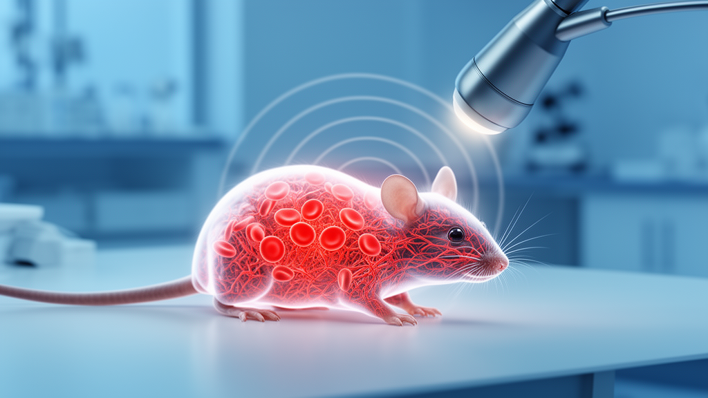
Researchers have shown that a contrast-free ultrasound technique called Super-resolution Ultrasound using Erythrocytes, or SURE, can visualise the microvasculature of subcutaneous tumours in mice without injecting any microbubble contrast agent. In a set of pilot studies published in Discover Imaging, the technique detected vessels approximately 30 micrometres in diameter, revealed vascular structures that conventional colour Doppler missed, and produced vascular density measurements that did not differ significantly from those obtained by ex vivo immunohistochemistry.
Tumour vascularity is an important biomarker of tumour growth and therapeutic response, but tumour-associated vessels are typically small in diameter, highly tortuous, and exhibit reduced and heterogeneous flow velocities. These features often place tumour microvasculature below the spatial and temporal resolution limits of current in vivo imaging modalities, so quantitative assessment of tumour vascularity remains challenging, and researchers frequently rely on surrogate measures such as gross tumour size or molecular tracer uptake.
SURE exploits the backscattering of red blood cells themselves rather than the localisation of injected microbubbles. It combines high-frame-rate imaging with specialised post-processing algorithms that amplify previously undetectable vascular signals, reducing the required data acquisition time to seconds while still enabling capillary-level imaging. The technique has previously been used to characterise microvasculature in the rat kidney and in the human lymph node, but the new study represents its pilot testing in preclinical oncology applications.
Three experiments were conducted using two murine tumour models. Pilot study 1 employed an LNCaP xenograft model in three mice, while pilot study 2 and the main study used C57BL/6 mice inoculated with the B16-F10 melanoma cell line, with two and five animals respectively. Tumours were established on the right flank and allowed to grow until approximately one centimetre in diameter before inclusion. Across the experiments, the team qualitatively compared the vascular imaging sensitivity and clinical applicability of SURE with colour Doppler imaging and ex vivo immunohistochemistry, and characterised optimal post-processing parameters for subcutaneous tumour imaging.
Imaging was performed with a 168-channel GE L8-18iD linear array transducer operated at a transmission frequency of 10 MHz, first connected to a commercial GE LOGIQ E9 scanner for B-mode and colour Doppler images, and then to a Verasonics Vantage 256 research system for SURE acquisition. The SURE sequence used pulse inversion with 24 emissions from 2 × 12 virtual sources, operating at a frame rate of 416.7 Hz, with recursive imaging raising the effective frame rate to 5000 Hz to facilitate erythrocyte tracking. Images were acquired over a one-minute period at an imaging depth limit of 2.5 cm. Both manually adjusted and automatically generated time-gain compensation settings were investigated, and all manual adjustments were completed by a single operator to reduce operator-induced bias.
Processing involved a windowed motion-correction method in which multiple low-motion data segments were manually selected from the one-minute acquisition and combined to produce approximately ten seconds of low-motion data. Singular value decomposition separated tissue, flow and noise, with upper and lower thresholds distinguishing flow from tissue and removing noise. The optimal singular value decomposition range and signal threshold varied between individuals and imaging planes, could not be predicted in advance, and required empirical screening of multiple threshold combinations for each scan. Processing was optimised for two distinct clinical objectives: high-sensitivity processing, maximising detection of small, low-flow vessels, and high clinical accuracy, minimising erroneous background signal. The team also introduced modifications to address clipping artefacts, including setting clipped values in the raw radiofrequency data to zero and refining element exclusion criteria.
The results showed a clear advantage over conventional Doppler in the melanoma model. In the B16-F10 tumours, SURE detected vascular signals in all acquired scans, and across all imaging planes it demonstrated comparable or improved vascular imaging compared with colour Doppler, revealing structures not detected with Doppler alone. In regions where flow was detected by both modalities, SURE provided enhanced spatial detail, enabling clearer delineation of vessel boundaries and branches, and allowed discrimination between small vessels in close proximity that were not resolvable in the corresponding colour Doppler images. High-sensitivity processing recovered substantially more tumour microvasculature than high-accuracy reconstruction but increased background signal in deeper image regions, while high-accuracy processing improved vessel contrast at the cost of detecting fewer low-intensity signals from small vessels.
Feasibility, however, differed markedly between the two tumour models. In the LNCaP xenograft model, flow was detected in only one of three tumours, and only when both colour Doppler and SURE were used; in the remaining two tumours, no flow was detected with either modality. All B16-F10 tumours, by contrast, showed detectable vasculature in every individual using both methods. The authors hypothesise that this reflects known differences in vascular architecture and haemodynamics between the models, noting that LNCaP tumours are characterised by lower vascular density and slower, often heterogeneous blood flow, which may impair the ability to identify vessels despite their structural presence, since SURE relies on distinguishing moving erythrocyte signals from stationary tissue.
SURE velocity processing also enabled identification of flow direction across the imaging plane and point-specific measurement of blood velocity and vessel diameter in vessels with sufficient signal strength, with nine to eighteen analysable vessels per high-accuracy scan. Across the five B16-F10 tumours in the main study, blood velocity measurements had a mean of 2.75 mm/s, with a median of 2.4 mm/s and a range of 1.2 to 7.5 mm/s, and varied across tumours, although the authors stress that these analyses should be interpreted as exploratory owing to the small sample size. Mean vessel lumen diameter was 121.31 micrometres, ranging from 52.8 to 251.1 micrometres, and estimated blood flow rates also differed between tumours. Comparable haemodynamic measurements could not be obtained using spectral Doppler, as no measurable flow was detected in any imaging planes. The authors caution that the volumetric flow estimates rely on simplified assumptions about vessel geometry and flow distribution, have not been independently validated against established flow measurement techniques in tumour tissue, and that two-dimensional super-resolution ultrasound measurements may underestimate flow velocity.
In the main study, quantitative vascular density was compared with ex vivo CD31 immunohistochemistry in five tumours, using fifteen measurements in total. SURE-derived vascular density had a mean of 4.42 percent, with a standard deviation of 2.33 percent and a range of 1.30 to 8.61 percent, while immunohistochemistry-derived measurements showed a mean of 3.00 percent, with a standard deviation of 0.66 percent and a range of 2.01 to 4.07 percent. Paired comparison using the Wilcoxon signed-rank test demonstrated no statistically significant differences between the two methods within any tumour. Qualitatively, however, fluorescent immunohistochemistry revealed a greater number of small vessels and finer vascular detail than SURE, a difference the authors attribute to the higher spatial resolution of ex vivo histological techniques and the absence of physiological motion and acoustic attenuation. They also note that SURE visualises perfused vessels containing flowing erythrocytes, whereas CD31 immunohistochemistry labels endothelial cells irrespective of perfusion status, so histology should be regarded as a complementary reference rather than a direct ground truth, particularly since no direct co-registration between ultrasound planes and histological sections was performed.
The study has acknowledged limitations. The sample size was small, reflecting the pilot nature of the work, the statistical analyses were underpowered, and all statistical findings should be considered exploratory. SURE images were affected by signal banding artefacts that persisted under both manual and automated time-gain compensation settings, believed to arise from strong reflectors such as air located outside the imaging plane, and substantial artefactual regions were manually excluded during vascular density analysis, although residual artefacts may have influenced quantitative measurements. SURE reconstruction remains sensitive to motion and operator-dependent parameter selection, since low-motion data segments were selected manually, and the impact of low-motion frame selection on image quality was not formally assessed. The comparison with colour Doppler should also be interpreted as a comparison with conventional Doppler ultrasound rather than the current state of the art, as the GE LOGIQ E9 system does not incorporate recent ultrafast Doppler approaches, and no formal quantitative comparison between the two modalities was performed.
Despite these constraints, the authors conclude that SURE represents a significant advance in contrast-free in vivo microvascular imaging, complementing rather than replacing colour Doppler by providing improved microvascular detail at the expense of increased acquisition and processing time. Compared with microbubble-based ultra-localised microscopy, SURE eliminates the need for contrast agent administration and permits substantially shorter acquisition times, although ultra-localised microscopy remains the more established super-resolution technique and may offer advantages in signal-to-noise ratio and recovery of microvascular density. Future work will focus on optimising acquisition and processing workflows, developing automated parameter selection and advanced motion correction strategies, and confirming the findings across varied tumour models, with the aim of enabling repeated, contrast-free imaging to monitor tumour progression and therapeutic response in preclinical oncology and, eventually, clinical practice.
Subject of Research: Contrast-free super-resolution ultrasound imaging of tumour microvasculature in murine subcutaneous tumour models
Article Title: Contrast-free super-resolution ultrasound imaging for microvascular assessment of murine subcutaneous tumour models in preclinical oncology
Article References: McDermott, A., Taghavi, I., Jimenez, N. P., Clausen, A. S., Naji, M. A., Kjær, A., Nielsen, M. B., Jensen, J. A., & Sørensen, C. M. (2026). Contrast-free super-resolution ultrasound imaging for microvascular assessment of murine subcutaneous tumour models in preclinical oncology. Discover Imaging, 3(1), Article 7. https://doi.org/10.1007/s44352-026-00028-3
Image Credits: AI Generated
DOI: 10.1007/s44352-026-00028-3
Keywords: super-resolution ultrasound, SURE, erythrocyte imaging, tumour microvasculature, preclinical oncology, murine tumour models, colour Doppler, vascular density, immunohistochemistry, B16-F10 melanoma, LNCaP xenograft, haemodynamics
News Source: Nathaniel Bowman. (October 28, 2026). Erythrocytes as contrast agents: ultrasound technique resolves tumour microvessels in mice. Scienmag.