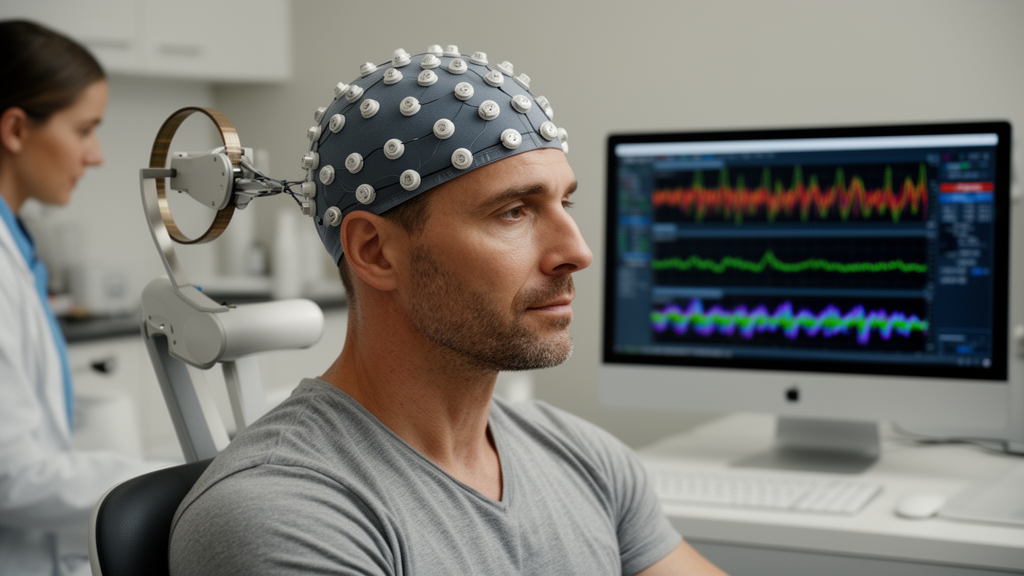
After twelve sessions, researchers documented changes in his resting brain wave patterns and a concurrent improvement on a computerized test of cognitive flexibility. The findings, published as a single-case study in Discover Neuroscience, are deliberately framed by their authors as preliminary observations of changes associated with the intervention rather than proof that the treatment works.
The case is notable because it illustrates an emerging strategy in neurorehabilitation: biomarker-informed neuromodulation, in which an individual’s measured neurophysiological abnormalities guide both where and how stimulation is delivered. Traumatic brain injury is a leading cause of persistent cognitive impairment in young, physically active populations, and it poses particular challenges for athletes, whose careers depend on rapid reactions, motor planning, and decision-making under pressure. Even when structural scans appear mild or focal, long-lasting functional alterations within large-scale brain networks can persist and undermine higher-order cognition, especially executive functions such as sustained attention, working memory, response inhibition, planning, and cognitive flexibility, which depend on distributed frontoparietal networks.
The participant’s injury followed a motor vehicle accident. Initial computed tomography imaging revealed a left frontotemporal subdural hematoma measuring 5.6 millimeters in thickness, a left frontal epidural hematoma measuring 6.7 millimeters, and mild cerebral contusion hemorrhage in the left frontoparietal region. Additional findings included a left parietal subperiosteal hematoma, a fracture of the right lateral pterygoid process, and signs of skull base trauma with air-fluid levels in the bilateral sphenoid sinuses. No midline shift was observed, indicating the absence of significant mass effect, and no surgical intervention was required. Vestibular assessment showed abnormalities consistent with bilateral vestibular dysfunction, including head-shaking nystagmus and impaired semicircular canal function.
Despite the absence of severe structural abnormalities, the man’s symptoms persisted. Two years after the injury he continued to report intermittent headaches, positional dizziness, impaired concentration, short-term memory difficulties, cognitive fatigue, attentional deficits, reduced working memory capacity, and balance instability during dynamic movements, a picture consistent with chronic post-traumatic cognitive and vestibular dysfunction. Conventional cognitive rehabilitation approaches, the authors note, have shown variable effectiveness and are often limited by prolonged intervention periods, dependence on active patient engagement, and heterogeneous outcomes, which has driven growing interest in non-invasive neuromodulation techniques such as transcranial magnetic stimulation as potential tools for modulating dysfunctional neural activity.
The intervention itself used the BetaOne C6 system, a low-intensity electromagnetic neuromodulation device registered for clinical use under Iranian medical device regulations. It generates magnetic fields with a maximum flux density of approximately 149.6 microtesla, far below the intensities used in conventional repetitive transcranial magnetic stimulation. The study defines low-intensity transcranial magnetic stimulation as the application of magnetic fields below the threshold required to directly depolarize cortical neurons or elicit motor-evoked potentials; rather than forcing neurons to fire, it is intended to modulate endogenous neuronal activity and neuroplasticity-related processes. Experimental work suggests such stimulation may influence neuronal excitability, synaptic plasticity, and functional network dynamics, although the precise biological mechanisms remain incompletely understood. Because the device employs proprietary stimulation algorithms, detailed engineering specifications such as pulse waveform and pulse width are not publicly available, and all sessions followed the manufacturer’s operating guidelines.
Protocol selection was driven by the participant’s baseline QEEG. Resting-state recordings were acquired under standardized conditions: five minutes with eyes closed, using a 19-channel active electrode montage positioned according to the international 10-20 system, sampled at 500 hertz with the Mitsar-202 system and Win-EEG software, electrode impedance below 5 kilohms, and the A1 electrode as reference. After artifact removal, at least 60 seconds of clean data were analyzed in NeuroGuide software across delta (1-4 Hz), theta (4-8 Hz), alpha (8-12 Hz), beta (12-25 Hz), sensorimotor rhythm (12-15 Hz), and high-beta bands, yielding absolute and relative power, hemispheric asymmetry, and connectivity measures that were transformed into age-adjusted Z-scores against a normative database. Based on the observed deviations, stimulation was sequentially applied to the Pz electrode site, chosen for posterior cortical oscillatory deviations, and to F3 and F4, which correspond approximately to the left and right dorsolateral prefrontal cortex, regions well established to participate in executive control, attentional regulation, working memory, and cognitive flexibility. Frequency-specific protocols from the device’s predefined library, spanning infra-low frequency through gamma and vagal protocols, were matched to the identified abnormalities.
The participant completed twelve sessions, each lasting twenty minutes, under standardized conditions. Tolerability and potential adverse events were monitored throughout, and no treatment-related adverse events or clinically significant side effects were reported. Cognitive performance was assessed before and after the intervention with the Intra-Extra Dimensional Set Shift task from the Cambridge Neuropsychological Test Automated Battery, a computerized test of attentional set formation, set-shifting, and cognitive flexibility in which the participant learns visual discrimination rules through trial-and-error feedback and must then shift attention between stimulus dimensions as demands change.
The results were descriptive, as befits a single-participant, quasi-experimental pre-post design with no control condition and no inferential statistical testing. On the cognitive side, Total Trials Adjusted fell from 59.70 at baseline to 53.48 after the intervention, and Total Errors Adjusted fell from 21.10 to 16.10. On the neurophysiological side, baseline QEEG had shown increased slow-wave activity in the delta and theta bands, alterations in frontal alpha and beta activity, and regional hemispheric asymmetry differences. After the intervention, comparisons revealed reductions in delta and theta power deviations at the Pz region, changes in alpha, beta, and high-beta activity in frontal regions including F3 and F4, and reduced departures from age-adjusted normative values in delta and theta on topographic Z-score maps. Connectivity analyses showed altered baseline patterns involving frontoparietal and frontotemporal regions, particularly within the right hemisphere, and post-intervention changes in intra- and inter-hemispheric connectivity measures, with several connectivity-related Z-score deviations appearing smaller relative to normative reference values.
The authors interpret these converging changes cautiously and consistently. They note that increased slow-wave activity after chronic traumatic brain injury has previously been associated with disrupted cortical regulation, reduced large-scale network efficiency, and increased cognitive demands during information processing, and that reductions in excessive slow-wave activity alongside more balanced oscillatory dynamics have been linked in prior neurophysiological investigations to more efficient excitatory-inhibitory regulation. The spatial distribution of the observed alterations corresponded with the individualized stimulation targets, consistent with the intended spatial targeting of the QEEG-guided approach. Yet they repeatedly emphasize that the electrophysiological changes are correlates associated with the intervention, not direct evidence of functional recovery, and that the concurrent behavioral improvements should be read as preliminary observations accompanying the neurophysiological changes rather than evidence that the intervention directly improved executive functioning. The study was not designed to establish causal relationships between the electrophysiological and cognitive outcomes.
The limitations are substantial and the authors enumerate them plainly. The single-case design precludes causal inference and severely limits generalizability; spontaneous recovery, placebo responses, or other non-specific effects cannot be excluded; the absence of a comprehensive standardized neuropsychological battery left other executive domains, including working memory, inhibitory control, planning, and problem solving, unassessed; and the lack of longitudinal follow-up means the durability of the observed changes is unknown. The authors call for randomized controlled designs with larger samples, comprehensive neuropsychological assessment batteries, multimodal outcome measures combining electrophysiology and neuropsychology, and long-term follow-up to determine the reproducibility, clinical relevance, and mechanisms of individualized QEEG-guided low-intensity stimulation. Within those boundaries, the case offers a concrete demonstration of what biomarker-informed neuromodulation looks like in practice: a protocol built from one patient’s measured oscillatory abnormalities, a documented pattern of post-intervention change across spectral power, asymmetry, and connectivity measures, and a parallel shift on a validated test of cognitive flexibility, all awaiting the controlled trials that will determine whether the approach holds clinical value for athletes and others living with chronic traumatic brain injury.
Subject of Research: A single-case study of QEEG-guided low-intensity transcranial magnetic stimulation in an athlete with chronic traumatic brain injury
Article Title: Effects of QEEG-guided low-intensity transcranial magnetic stimulation on executive network function in an athlete with chronic traumatic brain injury
Article References: Nasiri, A., Najafabadi, M. G., & Jahromi, M. A. (2026). Effects of QEEG-guided low-intensity transcranial magnetic stimulation on executive network function in an athlete with chronic traumatic brain injury. Discover Neuroscience, 21(1), Article 111. https://doi.org/10.1186/s13064-026-00330-0
Image Credits: AI Generated
DOI: 10.1186/s13064-026-00330-0
Keywords: traumatic brain injury, QEEG, transcranial magnetic stimulation, neuromodulation, executive function, cognitive flexibility, athletes, CANTAB, neurorehabilitation, case study, brain oscillations, Effects
News Source: Cassandra Pierce. (October 28, 2026). QEEG-Guided Low-Intensity Magnetic Stimulation Shows Preliminary Brain and Cognitive Changes in One Athlete With Chronic Traumatic Brain Injury. Scienmag.