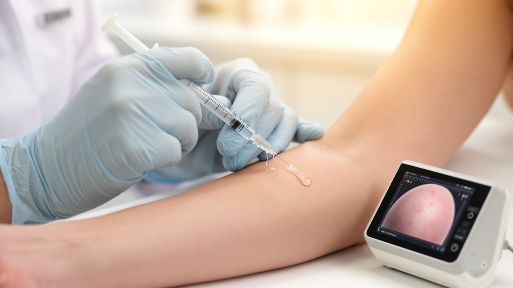
A liquid formulation of polycaprolactone, an injectable material better known in its microsphere-based filler form, has produced measurable changes in skin mechanical properties in a small prospective study of 30 adults, according to a report published in Dermatology and Therapy. Over six months of follow-up, participants who received three monthly superficial subcutaneous injections along the lateral periorbital and cheek regions showed significant reductions in maximum skin deformation and in the ratio of delayed to immediate deformation, alongside a decline in skin hydration and a pattern of mostly mild, self-limited adverse events. The investigators themselves caution, however, that the uncontrolled design and a late restriction of instrumental reporting mean the findings are exploratory rather than proof of efficacy.
The study, conducted at Siriraj with institutional review board approval on July 28, 2023, enrolled a convenience sample of 30 participants, 27 women and 3 men, with a mean age of 37.57 years and a range of 30 to 51. There was no formal sample-size calculation, no untreated comparison group, and no active comparator. All participants completed the three treatment sessions, with attendance of 29 at one month and 28 at both three and six months after the final treatment. The trial was registered retrospectively on the Thai Clinical Trials Registry on February 9, 2025, a limitation the authors acknowledge explicitly.
The intervention used GOURI, a liquid-form polycaprolactone product supplied as a 1-milliliter aqueous preparation containing 210 milligrams per milliliter of PCL, distilled water, and polyethylene glycol. Each session delivered 2 milliliters total, 1 milliliter per side, using a 23-gauge, 50-millimeter blunt cannula in a linear retrograde technique intended to place the material in the superficial subcutaneous plane immediately beneath the dermis. On each side, 0.1 milliliters was delivered along the crow’s-feet line and 0.3 milliliters along each of three cheek fanning tracks. Notably, the intended plane was assessed clinically only; no ultrasound or histology confirmed where the material actually deposited, a point the authors stress repeatedly when discussing mechanism.
The headline instrumental results come from probe-based skin biophysical measurements. Maximum skin deformation under suction, the Cutometer R0 parameter, fell from 0.153 plus or minus 0.062 millimeters at baseline to 0.098 plus or minus 0.041 millimeters one month after the second treatment, with a p value below 0.001, and remained significantly reduced at six months after the final session at 0.112 plus or minus 0.105 millimeters, with an overall p value below 0.001. Residual deformation after relaxation, R1, similarly declined from 0.078 plus or minus 0.036 millimeters to 0.058 plus or minus 0.034 millimeters at six months. Lower values on both parameters are conventionally interpreted as greater firmness and better elastic recovery, although the authors distinguish such statistical findings from clinically meaningful improvement, noting that no clinical response threshold was prespecified.
The corrected analysis of the delayed-to-immediate deformation ratio, R6, tells a more nuanced story. This dimensionless ratio, the quotient of delayed viscoelastic deformation to immediate elastic deformation, showed an overall change over time with an overall p value of 0.001. Mean R6 was 0.307 plus or minus 0.307 at baseline and fell to 0.174 plus or minus 0.277 at six months, with Bonferroni-adjusted comparisons against baseline reaching significance from before the third treatment through the six-month visit. The corrected results differ materially from values originally reported, and the authors emphasize that the large standard deviations relative to the means for R0 and R6 at six months indicate substantial heterogeneity among participants and do not support a claim of uniform, durable benefit.
Not every measured change pointed in a flattering direction. Skin hydration, measured with a Corneometer, actually decreased, from 55.28 plus or minus 19.54 at baseline to 46.23 plus or minus 14.31 at six months, a difference the authors say should be considered alongside the other outcomes without assuming it represents a beneficial effect. Sebum levels also declined, from 32.88 plus or minus 22.45 to 23.54 plus or minus 20.22. Melanin index decreased and the individual typology angle, an optical measure derived from color readings, increased over the follow-up period, but the authors note that pigmentary, sebum, and hydration measurements can all be influenced by season, sunscreen use, skincare habits, and environmental conditions, and transepidermal water loss and clinical barrier impairment were not assessed.
Safety findings were dominated by bruising and swelling. After the first treatment, bruising occurred in 6 of 30 participants, lasting a mean of about 16 days, with one case of swelling and one case of persistent postprocedural pain, all mild. After the second session, swelling occurred in 4 of 30 participants, three mild and one moderate, and after the third session in 3 of 30, all mild. All reported events resolved spontaneously without specific treatment, and no pruritus, nodules, granulomas, vascular complications, or serious adverse events appeared in the confirmed safety summary. Mean procedural pain scores on a 0-to-10 visual analog scale were 4.22, 4.76, and 4.95 after the three sessions, which the authors describe as appreciable despite topical and local anesthesia.
Participant-reported impressions were strikingly positive, but come with heavy caveats. At one month after the final treatment, 25 of 29 respondents, or 86.2 percent, reported marked or greatest improvement on the study’s percentage-improvement categories, and 23 of 28, or 82.1 percent, said the same at both three and six months. Yet the scale was nonvalidated, baseline photographs were not shown during assessment, and the ratings are explicitly labeled exploratory outcomes susceptible to recall and expectation effects. The representative clinical photographs were likewise not scored by blinded investigators and, as the authors put it, do not provide blinded evidence of improvement.
Two structural aspects of the report deserve particular attention. First, although the registry identifies Antera-based wrinkle indentation and texture as primary outcomes, this article is a focused, partial report that does not analyze those registered primary endpoints at all. Second, the original protocol included follow-up through 12 months, and the 9- and 12-month visits were completed, but calibration-related reliability concerns were identified for the Courage + Khazaka measurements at those late time points during review of the dataset. Because the concerns emerged after data collection and could not be resolved, the investigators submitted an institutional review board amendment, approved on May 21, 2025, restricting instrumental reporting to the baseline-through-six-month window. The authors acknowledge that this post-collection decision introduces a risk of selective time-point reporting and limits interpretation of durability, while noting the decision was based on measurement reliability rather than the direction or significance of the late results.
Mechanistic interpretation is similarly constrained. Preclinical work cited by the authors, including a UVB-exposed rat model in which intradermal liquid-form PCL delivered with a 34-gauge needle was associated with greater dermal thickness and type I collagen expression, cannot be transferred to the present 23-gauge cannula technique aimed at the superficial subcutaneous plane. Any dermal-level effect after such placement would have to arise indirectly, and the authors describe such mechanisms as hypothetical and unmeasured. They also decline to claim any advantage in tissue distribution or safety over conventional particulate PCL fillers, noting that the absence of delayed granulomas in a small series does not establish lower risk. Their conclusion is measured: skin mechanical and other biophysical changes were observed alongside predominantly mild, self-limited adverse events, but the uncontrolled design, the retrospective registration, and the restricted instrumental reporting mean these findings do not establish dermal deposition, neocollagenesis, comparative efficacy, or long-term safety, and larger controlled studies are needed.
Subject of Research: Skin mechanical changes and safety after liquid-form polycaprolactone injections for facial rejuvenation in a prospective single-arm study
Article Title: Skin Quality and Safety Following Liquid-Form Polycaprolactone for Facial Rejuvenation: A Prospective Single-Arm Study
Article References: Thongjaroensirikul, P., Wanitphakdeedecha, R., Maneeprasopchoke, P., Nokdhes, Y.-N., Buranaporn, P., Bhorntarakcharoen, W., Sittiwanaruk, S., & Techapichetvanich, T. (2026). Skin Quality and Safety Following Liquid-Form Polycaprolactone for Facial Rejuvenation: A Prospective Single-Arm Study. Dermatology and Therapy. https://doi.org/10.1007/s13555-026-01963-z
Image Credits: AI Generated
DOI: 10.1007/s13555-026-01963-z
Keywords: polycaprolactone, facial rejuvenation, skin quality, cutometer, single-arm study, adverse events, skin biomechanics, cosmetic dermatology, injectable biostimulator, prospective study, skin hydration, Skin
News Source: Ophelia Keating. (October 28, 2026). Liquid Polycaprolactone Injections Show Skin Firmness Signals in Small Uncontrolled Trial. Scienmag.