


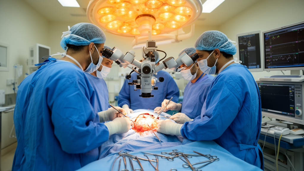
A ten-year review of more than 1,400 surgically treated brain and spinal cord tumors at Assiut University Hospital has revealed a distinctive epidemiological profile for Upper Egypt, with meningiomas emerging as the single most common tumor type and a striking U-shaped relationship between age and malignant behavior. The retrospective study, published in the Egyptian Journal of Neurosurgery, analyzed 1,404 histologically confirmed central nervous system tumors operated on between January 2011 and December 2020, making it one of the largest contemporary institutional series reported from the region.
The research team reviewed neurosurgery and pathology archives at Assiut University Hospital, which serves as the largest tertiary neurosurgical referral center in Upper Egypt and receives patients from multiple surrounding governorates. Only patients who underwent surgical resection with histopathological confirmation were included, and tumors were classified according to the 2016 World Health Organization Classification of Tumors of the Central Nervous System, with each lesion assigned International Classification of Diseases for Oncology, Third Edition topography and morphology codes. Cases managed conservatively or medically, such as prolactinomas treated with dopamine agonists, and lesions handled with stereotactic radiosurgery without biopsy, including small vestibular schwannomas and selected brainstem or optic pathway gliomas, were not captured by the design.
Of the 1,404 tumors analyzed, 1,333 were primary tumors and 71 were metastatic lesions. The mean patient age was 41.3 years with a standard deviation of 17.7 years, and the median age was 43 years, with individual cases ranging from six months to 85 years. Peak tumor occurrence fell in the fifth and sixth decades of life. Female patients accounted for 57.4 percent of the cohort, a proportion the authors attribute largely to the high frequency of meningioma among the women treated at the center. Meningioma was the most frequent tumor overall at 39.4 percent of all cases, followed by glioma at 27.7 percent, nerve sheath tumors at 8.7 percent, metastatic tumors at 5.0 percent, and pituitary adenoma, now also termed pituitary neuroendocrine tumor or PitNET, at 4.8 percent.
The pediatric and adult halves of the cohort told markedly different stories. Among patients aged 18 years or younger, gliomas represented the largest tumor group at 47.5 percent, followed by embryonal tumors at 28.2 percent and craniopharyngiomas at 7.2 percent. Pilocytic astrocytomas and ependymomas accounted for most pediatric gliomas, while medulloblastomas made up 80 percent of pediatric embryonal tumors. In adults, the picture reversed: meningiomas predominated at 44.8 percent, followed by gliomas at 24.9 percent and nerve sheath tumors at 9.7 percent. The differences between pediatric and adult patients were statistically significant, and the authors note that these patterns closely resemble observations from both earlier Egyptian studies and international series.
Anatomically, cranial tumors accounted for 82.4 percent of cases and spinal tumors for 17.6 percent. Among cranial tumors, meningioma was again the most common type at 40.2 percent, followed by glioma at 30.4 percent. Supratentorial lesions represented 79 percent of cranial tumors overall, but the split diverged sharply by age: in adults, 85 percent of cranial tumors were supratentorial, whereas in pediatric patients infratentorial lesions actually predominated, at 57.3 percent. In adults, meningiomas made up 49.7 percent of supratentorial tumors, followed by gliomas at 30 percent and pituitary adenomas at 7.6 percent. In children, gliomas led the supratentorial group at 50 percent, followed by craniopharyngiomas at 18.6 percent, while pediatric infratentorial tumors were split evenly between embryonal tumors and gliomas, each at 45.7 percent.
Spinal tumors showed their own internal architecture. They occurred most frequently in the dorsal region, which accounted for 56.7 percent of spinal cases, followed by the lumbar spine at 23.5 percent and the cervical spine at 18.6 percent. Intradural extramedullary tumors constituted the largest anatomical subgroup at 64.4 percent, with extradural tumors at 20.2 percent and intramedullary tumors at 15.4 percent. Meningiomas, at 36.3 percent, and nerve sheath tumors, at 30.3 percent, dominated the spinal tumor spectrum. Nerve sheath tumors were the most common lesions in the cervical and lumbar spine, while meningiomas led in the dorsal spine, where they accounted for 47.1 percent of tumors. Notably, gliomas represented 31 percent of lumbar spine tumors, and every one of these was a myxopapillary ependymoma.
Malignant tumors comprised 34.3 percent of the entire cohort, but that figure concealed a steep age gradient: malignancy affected 59.0 percent of pediatric patients compared with 30.7 percent of adults. Males constituted 56.2 percent of malignant cases, and glioblastoma was the single most common malignant entity, accounting for 28.4 percent of all malignant tumors in the series. To dissect the factors driving malignant behavior, the researchers performed multivariable logistic regression, categorizing age into pediatric, adult, and elderly groups with adults older than 18 but not older than 60 as the reference. When age was treated as a continuous variable, no significant association with malignancy emerged, but the categorical analysis exposed a non-linear, U-shaped pattern.
That regression analysis produced four independent predictors of malignancy. Pediatric patients had significantly higher odds of malignant tumor behavior than adults, with an adjusted odds ratio of 3.20 and a 95 percent confidence interval of 2.29 to 4.47. Elderly patients also carried elevated odds, with an odds ratio of 1.67 and a confidence interval of 1.20 to 2.32. Male sex remained independently associated with malignancy after adjustment for age and tumor site, with an odds ratio of 2.24 and a confidence interval of 1.78 to 2.82, and cranial location outperformed spinal location with an odds ratio of 1.89 and a confidence interval of 1.36 to 2.61. The authors interpret the U-shaped age pattern as biologically plausible, reflecting the differing histological composition of tumors across the lifespan: children are disproportionately affected by embryonal tumors and high-grade ependymomas, while elderly patients more frequently harbor glioblastomas and metastatic disease. They also caution that the surgical cohort reflects both underlying tumor biology and age-related surgical decision-making rather than a simple linear effect of aging.
The meningioma and glioma subanalyses added finer detail. Meningiomas occurred at a mean age of 47.6 years, with 98.9 percent of cases in adults and 72.7 percent in females. WHO grade I tumors accounted for 78.3 percent of meningiomas, and meningothelial meningioma was the most common histological subtype. Intracranial meningiomas made up 83.9 percent of the total, with the cerebral convexity the most frequent site at 41.1 percent, followed by parasagittal, sphenoid wing, olfactory groove, posterior fossa, and suprasellar locations; spinal meningiomas, at 16.1 percent, clustered overwhelmingly in the dorsal spine. Gliomas, meanwhile, were more common in males, who accounted for 57.2 percent of cases, in adults, and in intracranial sites. Astrocytic tumors comprised 79.7 percent of all gliomas, and glioblastoma was the dominant subtype, representing 35.1 percent of gliomas and 9.8 percent of all CNS tumors in the cohort. The mean age for glioblastoma patients was 49 years, compared with just 16.6 years for pilocytic astrocytoma, underscoring the heterogeneity within the glioma category.
The authors are explicit that their figures describe a surgical cohort, not population incidence. Because only resected, pathology-proven cases were included, tumors commonly managed without surgery, such as some pituitary tumors, optic pathway gliomas, germ cell tumors, and many metastatic lesions, are underrepresented, and the prominence of meningioma likely reflects case ascertainment in addition to true disease frequency. The absence of routine molecular testing also meant that integrated diagnoses under the 2021 WHO CNS5 classification could not be assigned, and historical terms such as primitive neuroectodermal tumor and diffuse astrocytoma may not map directly onto current categories. As a single-center study, the findings reflect operative decision-making and referral patterns at a large tertiary center rather than the true disease burden of the Egyptian population. Even so, the scale of the series and the hospital’s broad referral base offer a valuable window into the pattern of surgically managed CNS tumors in Upper Egypt, and the authors call for future multicenter studies incorporating molecular classification and capturing non-surgical cases to define that burden more completely.
Subject of Research: The histopathological spectrum and predictors of malignancy among surgically treated central nervous system tumors at a tertiary hospital in Upper Egypt over ten years.
Article Title: Histopathological spectrum and clinicopathological profile of surgically treated central nervous system tumors at Assiut University Hospital: a 10-year retrospective study
Article References: Mohammad, F., Abd Elhakeem, A. A. E., El-Gazzar, A. F., & Essa, A. A. (2026). Histopathological spectrum and clinicopathological profile of surgically treated central nervous system tumors at Assiut University Hospital: a 10-year retrospective study. Egyptian Journal of Neurosurgery, 41(1), Article 178. https://doi.org/10.1186/s41984-026-00688-7
Image Credits: AI Generated
DOI: 10.1186/s41984-026-00688-7
Keywords: CNS tumors, meningioma, glioma, glioblastoma, medulloblastoma, Upper Egypt, retrospective study, WHO classification, pediatric tumors, malignancy predictors, spinal tumors, tertiary referral center
News Source: Cassandra Pierce. (October 28, 2026). Meningiomas Dominate a Decade of Brain Surgery in Upper Egypt. Scienmag.



