The condition, a chronic immune-mediated fibro-inflammatory disorder that can masquerade as meningioma, schwannoma or ordinary inflammation, was tracked in nine biopsy-proven cases diagnosed at the Neuropathology Laboratory of the All India Institute of Medical Sciences in New Delhi between January 2021 and December 2024. The study, published in Discover Neuroscience, integrates clinical, serological, radiological, pathological, treatment and follow-up data for every patient, and its central message is sobering: not one of the nine lesions was correctly identified as IgG4-related disease before tissue examination in all but a single case.
The cohort was strikingly young. The mean age was 25.9 years with a standard deviation of 8.8 years, and patients ranged from 11 to 41 years old, with men outnumbering women two to one. That profile contrasts with the global literature, which typically describes onset in the fifth to sixth decades of life. The authors caution that their cases were ascertained through a tertiary neuropathology archive of surgically sampled lesions, which may preferentially capture younger patients with focal, mass-forming or diagnostically uncertain disease, so the age difference should be interpreted with that selection in mind. Still, they raise the possibility that ethnic or environmental modifiers influence the expression of isolated central nervous system disease in Indian populations, a question that only broader regional datasets can answer.
Headache was the most common presenting symptom, affecting five of the nine patients, followed by seizures in three. The location of disease shaped the clinical picture. The frontal or frontotemporal dura was most frequently involved, in three cases, with the cerebellopontine angle dura involved in two. The remaining lesions occupied the right occipital and cerebellar dura, the tentorium cerebri, the inferior cerebellum and the right parietal extra-axial convexity. Notable presentations included a tentorial lesion causing headache, visual loss and diplopia; an inferior cerebellar lesion producing gait ataxia; and a cerebellopontine angle lesion associated with quadriplegia and bilateral hearing loss. The diversity of these syndromes underscores how anatomical spread, rather than any disease-specific signature, drives what patients actually experience.
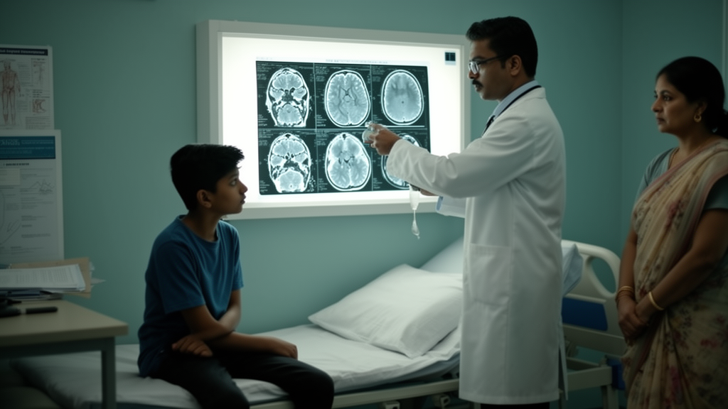
Radiology added to the diagnostic confusion rather than resolving it. The most frequent imaging impression was hypertrophic pachymeningitis or non-specific inflammatory disease, in four of nine cases, followed by lesions suggestive of meningioma in three. One lesion was read as a vestibular schwannoma at the left cerebellopontine angle. Only a single case was flagged radiologically as possible IgG4-related disease before surgery. The authors argue that this near-total failure of preoperative imaging recognition makes a strong case for placing IgG4-related disease on the differential diagnosis list for atypical or inflammatory dural lesions, particularly when conventional causes such as infection or meningioma have been excluded.
Serological testing told a similarly incomplete story. Preoperative serum IgG4 levels were available for only four of the nine patients, with values of 1.1, 1.4, 1.7 and 4.3 grams per litre, and three of those four exceeded the 1.35 grams per litre threshold conventionally used to define elevation. The mean of the measured values was 2.47 grams per litre with a standard deviation of 1.6. Cerebrospinal fluid analysis, performed in three cases, showed raised protein in two with normal glucose, and total leukocyte counts of five to ten lymphocytes per cubic millimetre in all three. Erythrocyte sedimentation rate, available in five cases, was raised in all, ranging from 11 to 66 millimetres per hour, while rheumatoid factor, antinuclear antibody and antineutrophil cytoplasmic antibodies, tested in three cases, were uniformly negative. The authors note that testing for inflammatory and vasculitic mimics was not performed uniformly, so only documented results were considered.
Histopathology remained the decisive arbiter. Every lesion exhibited dense lymphoplasmacytic inflammation with storiform fibrosis, predominantly involving the dura and extending into the meninges and superficial brain parenchyma. The infiltrate was polymorphous, containing plasma cells, mature lymphocytes, histiocytes and occasional eosinophils. Obliterative phlebitis, one of the three classic features of the disease, appeared in only two of the nine cases, which the authors describe as an uncommon but diagnostically supportive finding in this cohort and possibly a site-related feature of intracranial disease. Immunophenotyping in all cases showed more than ten IgG4-positive plasma cells per high-power field and an IgG4-to-IgG-positive plasma cell ratio exceeding 40 percent, quantified in hot spots of highest density. No atypia, mitotic activity, necrosis, granulomas, emperipolesis, giant cells or fungal elements were identified, and ancillary immunostaining including EMA, CD3, CD20, histiocyte markers and CD138 helped exclude lymphoplasmacyte-rich meningioma, inflammatory myofibroblastic tumour, plasmacytoma, lymphoma and histiocytic disorders such as Rosai-Dorfman and Erdheim-Chester disease.
Classification followed the 2020 revised comprehensive diagnostic criteria for IgG4-related disease. Three patients with elevated serum IgG4 were classified as definite cases, while the remaining six, who met the clinical, radiological and pathological domains but had not been tested for serum IgG4, were classified as probable. The authors stress that serum IgG4 may be normal in organ-limited disease and cannot independently establish or exclude the diagnosis, a point of particular consequence in resource-limited settings where the test is not routinely available. In such contexts, they argue, the integration of tissue morphology, immunophenotyping and radiological correlation becomes the practical pathway to diagnosis, and the pathological features of storiform fibrosis with a quantitative IgG4 plasma cell threshold are critical to avoid misdiagnosis and unnecessary surgery.
Treatment outcomes were uniformly favourable in this cohort. All patients received corticosteroid-based therapy, with or without adjunctive immunosuppressive agents, and all responded. The three patients with elevated serum IgG4 showed post-therapy reductions to 1.2, 0.72 and 0.9 grams per litre respectively. No patient died of the disease, and no clinical recurrence or radiological progression was documented during a mean follow-up of 30.58 months with a standard deviation of 10.63. The authors interpret this as evidence of a favourable disease trajectory with prompt immunosuppression and of the reversibility of the inflammatory phase when recognised early, while recommending longer clinical observation. They also note that in the broader literature, glucocorticoids effectively reduce inflammation but relapses are common, often necessitating long-term management with steroid-sparing agents such as rituximab, and that surgery is generally reserved for isolated compressive lesions given the risks of operating near critical nervous system structures.
Perhaps the most distinctive observation of the series is that every case showed isolated central nervous system involvement, with no extra-CNS or systemic disease documented in the available records. That finding is less frequently reported elsewhere, where multiorgan disease predominates and extra-CNS manifestations occur in roughly half of patients. The authors attribute this partly to referral bias at tertiary neurosurgical centres but argue it also highlights the need to recognise CNS IgG4-RD as a potential organ-limited immune disorder at presentation. They acknowledge that the pathogenesis of the disease is not fully elucidated, though aberrant interactions between CD4-positive T cells and B cells are thought to drive the characteristic histopathological changes. Elevated serum IgG4 typically accounts for less than six percent of total immunoglobulins, and up to one-third of patients in the wider literature may present with normal concentrations, making isolated CNS disease especially challenging.
The study’s authors present the series as the largest and first comprehensive regional dataset highlighting isolated CNS involvement in IgG4-related disease, and its practical implications are directed at clinicians confronting dural masses and pachymeningitis of uncertain cause. A multidisciplinary diagnostic approach, they conclude, is essential for accurate evaluation, may facilitate timely diagnosis and could help avoid unnecessary surgical interventions in this potentially reversible immune-mediated condition.
Subject of Research: IgG4-related disease of the central nervous system in a retrospective Indian case series
Article Title: Clinicopathologic spectrum and diagnostic insights from IgG4-related disease of the CNS
Article References: Jain, S., Jangir, H., Shah, S. A., Roy, C., Suri, V., Garg, A., Sharma, M. C., Kumar, A., Mishra, S., Verma, S., Gurjar, H. K., Sharma, R., & Phalak, M. (2026). Clinicopathologic spectrum and diagnostic insights from IgG4-related disease of the CNS. Discover Neuroscience, 21(1), Article 113. https://doi.org/10.1186/s13064-026-00343-9
Image Credits: AI Generated
DOI: 10.1186/s13064-026-00343-9
Keywords: IgG4-related disease, central nervous system, hypertrophic pachymeningitis, dural masses, serum IgG4, histopathology, immunohistochemistry, corticosteroid therapy, retrospective cohort, India, neuropathology, diagnostic criteria
News Source: Ophelia Keating. (October 28, 2026). Rare immune disease of the brain lining misdiagnosed as tumours in young Indian patients. Scienmag.