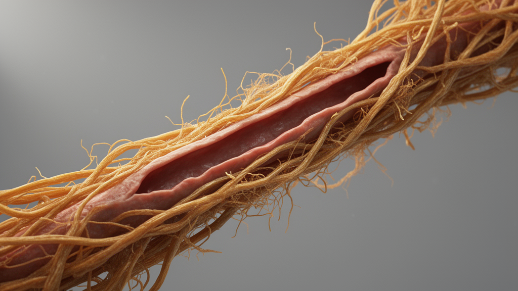
The extracellular matrix, long dismissed as little more than the biological scaffolding that holds blood vessels together, is now being recast as an active participant in vascular aging. Their synthesis positions the aged matrix as both a driver and a promising intervention target for cardiovascular aging and disease.
The review, published on 19 October 2026 as an open-access article in volume 32 of the journal, builds on a conceptual shift that has gathered momentum over the past two decades. For much of modern vascular biology, the extracellular matrix was treated as a passive scaffold that preserved vessel architecture. The authors contend that this view is outdated. The matrix, a dynamic network of collagens, elastin, proteoglycans, glycosaminoglycans, fibronectin, laminins and matricellular proteins, is continuously synthesized, modified and reorganized, and it regulates the bioavailability of growth factors and cytokines while controlling cell adhesion, migration, differentiation, survival and inflammatory state.
What changes with age is not simply the quantity of matrix material but its character. The authors identify a set of interconnected hallmarks of the aged vascular matrix. Collagen accumulates and becomes excessively crosslinked, driven in part by the enzyme lysyl oxidase and by non-enzymatic glycation that produces advanced glycation end products. Elastin, the remarkably durable protein that allows arteries to expand and recoil with each heartbeat, fragments irreversibly with age, because mature elastin fibers have an extremely long half-life and regeneration in adulthood is limited. The result is progressive matrix stiffening: aged arteries become more rigid, with lower compliance and a reduced ability to adjust to blood flow pulsations.
These structural changes carry biochemical consequences. As elastin degrades, proteases such as matrix metalloproteinases cleave it into short peptide fragments known as matrikines, including elastin-derived peptides bearing the conserved GxxPG/VGVAPG motif. These fragments bind the elastin receptor complex and trigger signaling cascades through neuraminidase-1, regulating calcium signaling, extracellular signal-regulated kinase activation, cytokine production, cellular migration and inflammatory responses in endothelial cells, vascular smooth muscle cells, fibroblasts and immune cells. Recent studies in humans and mice report high circulating levels of elastin, fibronectin and hyaluronic acid-derived fragments with age, and the elastin-derived peptides show the strongest association with aging phenotypes. In mice, chronic administration of elastin-derived peptides reduced lifespan and physical capacity and elicited senescence, systemic inflammation and obesity.
The mechanistic core of the review concerns how vascular cells sense their changing matrix environment. Cells continuously interpret matrix-derived cues through integrins, discoidin domain receptors, syndecans and mechanosensitive ion channels such as Piezo1 and TRPV4. Increased matrix stiffness facilitates force transmission through integrins and sustains activation of focal adhesion kinase and Src family kinases, while mechanical forces at focal adhesions are relayed through RhoA/ROCK-dependent cytoskeletal remodeling. Among the most consequential downstream mediators are the transcriptional co-activators YAP and TAZ: under compliant matrix conditions they remain largely cytoplasmic and inactive, but stiff matrices promote their nuclear translocation, where they interact with TEAD transcription factors to regulate genes involved in proliferation, fibrosis, inflammation and cellular plasticity. Increased YAP/TAZ activity has been implicated in vascular remodeling, atherosclerosis, fibrosis and vascular calcification.
As the matrix ages, these mechanosensitive pathways become chronically activated, and the review argues that this persistent signaling is what converts structural damage into cellular reprogramming. Sustained mechanotransduction drives endothelial dysfunction, a phenotypic switch in vascular smooth muscle cells from a contractile to a synthetic state, fibroblast activation, inflammation, calcification and the induction of senescence. The authors frame this as a transition from the matrix as structure to the matrisome as information: in healthy vessels, matrix architecture encodes tissue identity and preserves cellular specialization, whereas in aged vessels, stiffened, glycated and disorganized matrix information becomes pathogenic, biasing cell behavior toward maladaptive states.
A striking feature of the review is its treatment of the relationship between matrix aging and cellular senescence as bidirectional and self-amplifying. Senescent vascular cells, marked by persistent cell cycle arrest, elevated p16, p21 and p53, and a senescence-associated secretory phenotype rich in cytokines, chemokines and matrix proteins, profoundly reshape the matrix they inhabit. Evidence cited includes increased fibronectin in replicatively exhausted human endothelial cells, elevated matrix metalloproteinase-2 and fibronectin in aged rat aortas, and single-cell RNA-sequencing of young and old mouse aortas showing that elevated CXCL12 in senescent endothelial cells partially accounts for impaired endothelial function in aged mice, an impairment ameliorated by fisetin supplementation. In the media of the vessel wall, senescent vascular smooth muscle cells secrete interleukin-1 alpha, which drives secretion of active matrix metalloproteinase-9 and reduced collagen deposition, and immunohistochemistry of aortas from young and old patients revealed high SOX9 levels in p16-positive smooth muscle cells, where SOX9 overexpression promoted senescence and a shift toward an osteo/chondrogenic phenotype.
The reverse direction is equally consequential: the aged matrix can itself initiate and maintain senescence. Mice carrying collagenase-resistant collagen type I alpha 1 mutations developed features of premature aging, including age-related hypertension and increased aortic collagen, and culturing human vascular smooth muscle cells on collagen from these mice accelerated replicative senescence. Knockdown of lumican ameliorated angiotensin II-induced senescence in smooth muscle cells in vitro, accompanied by lower collagen deposition and reduced vascular aging in mice. Increased matrix stiffening has also been reported to contribute to smooth muscle cell senescence by impairing mitochondrial-endoplasmic reticulum contact in abdominal aortic aneurysm, an effect ameliorated by atorvastatin. Notably, a comprehensive proteomic profiling across multiple human tissues found the aorta to be the most susceptible tissue to aging, with age-related protein changes accumulating from as early as 30 years of age and surpassing other tissues by 45, leading the authors to suggest that arteries may act as a senohub and a source of systemic aging.
Oxidative stress threads through the entire framework as an additional effector. Age-associated reactive oxygen species from mitochondria and NADPH oxidase modify matrix composition and integrity, and advanced glycation end products act as ligands for receptors such as RAGE, triggering Rho signaling in endothelial cells and NADPH oxidase-driven, NF-kappaB inflammatory signaling in vascular smooth muscle cells. This creates a feed-forward loop in which oxidative stress promotes glycation and matrix remodeling, while the aged, stiff matrix in turn boosts oxidase activity, mitochondrial dysfunction and further reactive oxygen species generation. Experimental support spans antioxidant administration reducing collagen deposition in rat hearts, superoxide dismutase treatment attenuating fibronectin levels in diabetic rats, and antioxidant or hydrogen peroxide scavenger treatment reducing vascular stiffness and elastin alterations in hypertensive rodents.
The authors are careful about the limits of the current evidence. Distinguishing whether matrix alterations drive cellular dysfunction or result from it remains, by their own account, a central unresolved challenge, and they call for experimental systems, including tissue-engineered matrices, hydrogels and decellularized scaffolds, capable of independently manipulating matrix composition, stiffness or organization while controlling cellular age. Whether matrikines act primarily as biomarkers of tissue damage, endocrine-like signaling molecules or mediators of inter-organ communication remains largely unknown, and whether aged vascular cells placed in a youthful extracellular environment can rescue youthful phenotypes is an open question. Still, the therapeutic implications are considerable: if the aging matrix is an upstream driver rather than a downstream consequence, interventions aimed at preserving matrix integrity, reducing pathological crosslinking, modulating mechanotransduction, eliminating senescent cells or restoring youthful matrix composition could offer strategies that simultaneously influence inflammation, fibrosis, calcification and senescence, reframing the extracellular environment itself as a target for extending cardiovascular resilience.
Subject of Research: The role of the aging extracellular matrix in driving vascular cell reprogramming and cardiovascular aging
Article Title: The Aging Extracellular Matrix as a Driver of Vascular Reprogramming
Article References: Shalash, R., Tsitsipatis, D., & Herman, A. B. (2026). The Aging Extracellular Matrix as a Driver of Vascular Reprogramming. Artery Research, 32(1), Article 45. https://doi.org/10.1007/s44200-026-00144-9
Image Credits: AI Generated
DOI: 10.1007/s44200-026-00144-9
Keywords: extracellular matrix, vascular aging, arterial stiffness, collagen crosslinking, elastin fragmentation, mechanotransduction, cellular senescence, YAP/TAZ signaling, matrikines, advanced glycation end products, oxidative stress, cardiovascular disease
News Source: Beatrice Stafford. (October 27, 2026). Aging Extracellular Matrix Emerges as Active Driver of Vascular Aging. Scienmag.