


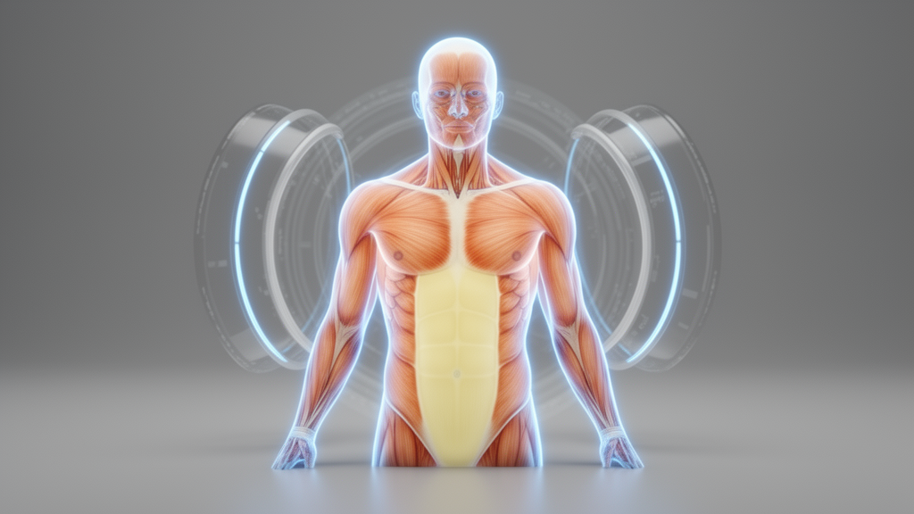
Artificial intelligence can measure the muscle and fat distributed throughout a patient’s body just as reliably from a whole-body MRI scan as from a low-dose CT scan acquired on the very same day, according to a new study of patients with multiple myeloma. But the study also delivers a cautionary note: while the two modalities track each other closely, they do not produce identical numbers. MRI consistently returned lower estimates for skeletal muscle and visceral fat than CT, a systematic bias that the authors say must be corrected before measurements from the two technologies can be pooled or compared in clinical practice.
The research, published in the journal Cancer Imaging, addresses a growing problem in oncology. Changes in body composition, particularly the loss of skeletal muscle and the redistribution of adipose tissue, have been linked to chemotherapy intolerance, increased postoperative complications and reduced survival in patients with cancer. Imaging-derived body composition measures are therefore increasingly viewed as potential prognostic biomarkers that could complement conventional clinical factors. Yet the traditional way of measuring them, a manual or semi-automatic two-dimensional slice, most often taken at the level of the third lumbar vertebra, is limited by slice selection bias, operator dependence and its inability to capture whole-body complexity.
Deep learning has changed that calculus. Automated segmentation algorithms such as TotalSegmentator can now delineate skeletal muscle, subcutaneous adipose tissue and abdominal visceral adipose tissue across entire imaging volumes rapidly and reproducibly. What remained uncertain, the authors argue, was whether these fully automated, volumetric measurements would agree across imaging modalities. Previous comparative studies had reported good correlations between CT- and MRI-derived metrics, but those investigations were largely restricted to single-slice or region-of-interest methods and often relied on manual processing. According to the study, no direct MRI-CT comparison had previously been published for whole-body volumetric body composition analysis.
To fill that gap, the researchers retrospectively analyzed data from consecutive patients with newly diagnosed multiple myeloma enrolled in the prospective GMMG-HD7 and GMMG-HD8/DSMM-XIX trials. Of 123 consecutive patients who had undergone baseline fluorine-18 FDG PET-CT at one of two large oncologic imaging centers between October 2018 and February 2025, 81 were ineligible because they had not received a whole-body MRI on the same day. That left 42 patients, imaged between March 2019 and February 2025, whose same-day PET/CT and whole-body MRI examinations formed the study cohort. Twenty of the 42 patients were female, the mean age was 59.9 years with a standard deviation of 6.9 years, and the mean body mass index was 26.4 kilograms per square meter.
The imaging protocols reflected routine clinical practice. Whole-body MRI was performed on a 1.5 Tesla system without intravenous contrast, and the volumetric analysis was based on the coronal T1-weighted turbo spin-echo sequence, using the vendor’s automatically composed whole-body image. The CT component came from the low-dose CT scans, acquired at 120 kilovoltage peak and 30 effective milliampere-seconds, that are routinely obtained for attenuation correction and anatomical co-registration during PET/CT. The automated pipeline, built on TotalSegmentator for CT and its MRI variant for magnetic resonance images, segmented skeletal muscle, subcutaneous fat and abdominal visceral fat in both modalities. To ensure comparability across patients with differing scan coverage, quantitative analyses were restricted to regions above the tibial plateau, and the ninth thoracic vertebra served as the boundary defining the abdominal visceral fat compartment.
The results showed strikingly high agreement for whole-body volumetric metrics. Intraclass correlation coefficients exceeded 0.95 for all three tissues: 0.99 for subcutaneous adipose tissue, 0.98 for skeletal muscle and 0.99 for visceral adipose tissue, with Pearson correlations of 0.98 to 0.99, all statistically significant. But Bland-Altman analysis, which examines the differences between paired measurements rather than merely their correlation, exposed systematic modality-related biases. Skeletal muscle volumes were on average 2,582 milliliters lower when extracted from MRI than from CT, and visceral fat volumes were 211 milliliters lower, both differences highly statistically significant. Subcutaneous fat showed a smaller, non-significant difference of 263 milliliters.
For single-slice, two-dimensional analysis at the third lumbar vertebra, agreement remained high for the fat compartments, with intraclass correlation coefficients of 0.97 for subcutaneous fat and 0.98 for visceral fat, but dropped to 0.87 for skeletal muscle, with a Pearson correlation of 0.77. Again, Bland-Altman plots revealed that skeletal muscle was systematically and significantly lower when measured from MRI, with a mean bias of 18.4 square centimeters. The fat compartments also trended lower on MRI, by 5.4 and 3.4 square centimeters respectively, but those differences did not reach statistical significance. The pattern suggests that the muscle compartment, with its complex anatomy and susceptibility to partial volume and motion effects, is where modality differences concentrate most.
To determine whether segmentation errors could explain the discrepancies, a board-certified radiologist with more than six years of experience visually reviewed every segmentation on a five-point quality scale. More than 95 percent of segmentations were rated as sufficient for both modalities across all compartments, with mean scores between 3.7 and 3.9. The identified failure modes were few but instructive. On CT, artifacts led to abdominal organs being misclassified as visceral fat, and cervical vascular structures were occasionally labeled as muscle. On MRI, visceral fat segmentation was sometimes missing, possibly due to bowel motion and partial volume artifacts, cervical subcutaneous fat was occasionally missed, and muscle boundaries were incompletely delineated. Notably, a large myeloma-typical focal bone marrow lesion, which replaces fatty marrow with soft tissue mimicking the density and signal intensity of muscle, was misclassified as skeletal muscle on both CT and MRI in one patient.
The authors suggest several explanations for the residual intermodal differences. Minor segmentation inaccuracies at anatomical borders likely reflect partial volume effects and motion artifacts inherent to MRI signal characteristics, which challenge human readers and deep learning models alike. The non-contrast CT protocol and possible underrepresentation of cervical vascular anatomy in training data may have contributed to the misclassification of vessels as muscle. In addition, because the analysis extended to the extremities, regional differences in scan coverage, particularly the upper arms sometimes only partially captured within the whole-body MRI field of view, may have introduced further discrepancies, even though all scans were standardized from head to tibia.
The study’s conclusions carry practical weight for a field moving toward multimodal imaging. As MRI increasingly assumes a central role in the diagnosis and treatment monitoring of multiple myeloma, the authors note that AI-based body composition analysis from whole-body MRI would yield results comparable to those known from the PET/CT literature, provided that the observed modality-specific biases are accounted for. The findings are limited to one widely used MRI protocol, one scanner field strength and the TotalSegmentator-based segmentation models, so performance of other models or sequences may vary. The authors call for future validation across diverse MRI protocols, reconstruction kernels and deep learning models, and for prospective assessment of clinical integration. For now, the message is clear: automated body composition metrics from CT and MRI are highly concordant, but the values are not directly interchangeable without correcting for consistent, tissue-specific biases, particularly for skeletal muscle and visceral fat.
Subject of Research: Intermodal agreement of AI-based automated body composition analysis between whole-body MRI and low-dose PET/CT in multiple myeloma patients
Article Title: AI-based automated body composition analysis from whole-body MRI and (PET-) CT: an intermodal comparison from same-day examinations
Article References: Holzschuh, J. C., Rotkopf, L. T., Neelsen, C., Bauer, F., Zhang, K. S., Menis, E., Hajiyianni, M., Mai, E. K., Weinhold, N., John, L., Sachpekidis, C., Dimitrakopoulou-Strauss, A., Raab, M.-S., Goldschmidt, H., Jende, J., Delorme, S., Schlemmer, H.-P., & Wennmann, M. (2026). AI-based automated body composition analysis from whole-body MRI and (PET-) CT: an intermodal comparison from same-day examinations. Cancer Imaging, 26(1), Article 149. https://doi.org/10.1186/s40644-026-01134-5
Image Credits: AI Generated
DOI: 10.1186/s40644-026-01134-5
Keywords: artificial intelligence, deep learning, body composition, whole-body MRI, PET/CT, skeletal muscle, visceral adipose tissue, multiple myeloma, TotalSegmentator, image segmentation, Bland-Altman analysis, oncology imaging
News Source: Nathaniel Bowman. (October 27, 2026). AI body composition scans agree across MRI and CT but reveal systematic biases. Scienmag.



