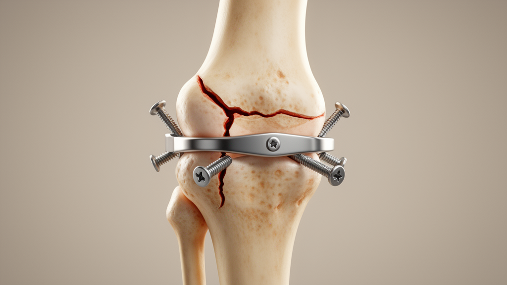
A finite element study comparing four ways of fixing complex tibial plateau fractures has found that combining a lateral plate with an intramedullary nail produced the least displacement and the lowest peak implant stress under the simulated loading conditions. The modelling work, published in BMC Musculoskeletal Disorders, simulated AO/OTA type 41-C2 tibial plateau fractures and evaluated lateral plate fixation, intramedullary nail fixation, dual-plate fixation and a combined plate-nail construct.
Four fixation strategies were simulated in the engineering software Abaqus: Group A used lateral plate fixation alone, Group B used an intramedullary nail, Group C combined lateral and medial plates in a dual-plate construct, and Group D paired a lateral plate with an intramedullary nail.
Two loading conditions were applied to each construct. The team compared maximum displacement, apparent construct stiffness and von Mises stress distribution, standard measures of how much a construct deforms and where stress concentrates in the implants.
Under axial loading, maximum displacement occurred at the posteromedial tibial plateau in all four groups. The lateral plate construct displaced 1.421 millimetres, the intramedullary nail 0.315 millimetres, the dual-plate construct 0.816 millimetres and the plate-nail combination 0.308 millimetres. Peak von Mises stresses in the fixation systems followed the same ordering: 630.7 megapascals for the lateral plate, 134.0 for the nail, 380.1 for the dual plates and 123.3 for the plate-nail construct.
Under varus loading the pattern shifted in location but not in ranking. Maximum displacement occurred at the lateral plateau in the lateral plate and intramedullary nail groups, reaching 2.598 and 0.622 millimetres respectively, and at the medial plateau in the dual-plate and plate-nail groups, at 1.136 and 0.335 millimetres. Peak implant stresses were 755.1 megapascals for the lateral plate, 88.0 for the nail, 317.3 for dual plates and 72.8 for the plate-nail construct.
Across both loading scenarios, the plate-nail combination showed the lowest maximum displacement and the lowest peak implant stress of the four simulated constructs, while the lateral plate alone showed the highest values on both measures. The intramedullary nail performed close to the plate-nail construct, and the dual-plate configuration fell in between. The authors conclude that, under the specific conditions simulated, intramedullary nail fixation and plate-nail fixation showed lower displacement and peak implant stress than lateral plate and dual-plate fixation.
The findings are strictly biomechanical. In this study those inputs came from a single healthy young male tibia and two idealised loading cases, so the model does not capture variation in bone quality, patient anatomy, surgical accuracy or the complex, repetitive and dynamic loads of real weight bearing.
The authors position the work as comparative biomechanical evidence to inform further evaluation of these fixation constructs, not as proof of clinical superiority. The authors position the work as comparative biomechanical evidence to inform further evaluation of these fixation constructs.
Subject of Research: Finite element comparison of four internal fixation strategies for tibial plateau fractures
Article Title: Finite element analysis of four different internal fixation strategies for tibial plateau fractures
Article References: Xie, L., Zhu, L., Chen, J., Luo, D., Zhou, L., Lian, K., Xie, W., & Liu, H. (2026). Finite element analysis of four different internal fixation strategies for tibial plateau fractures. BMC Musculoskeletal Disorders. https://doi.org/10.1186/s12891-026-10667-6
Image Credits: AI Generated
DOI: 10.1186/s12891-026-10667-6
Keywords: tibial plateau fracture, finite element analysis, internal fixation, intramedullary nail, dual-plate fixation, biomechanics, orthopaedic surgery, von Mises stress, AO/OTA 41-C2, plate-nail fixation, Finite, element
News Source: Ophelia Keating. (October 27, 2026). Plate-nail fixation shows lowest stress in simulated tibial plateau fractures. Scienmag.