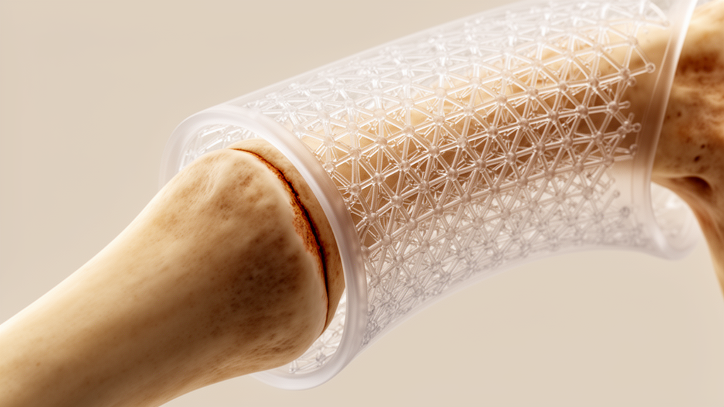
Engineers at Colorado State University have shown that a 3D-printed polycaprolactone sleeve wrapped around a highly porous bone scaffold can significantly change the mechanical environment inside a large bone defect. In tests on ovine cadaveric metatarsal bones fitted with a critical-sized segmental defect, the sleeve reduced transverse strain in the scaffold by 8.2 percent, bringing it down to approximately 0.03 percent, while leaving axial strain essentially unchanged. The findings, published in Frontiers in Bioengineering and Biotechnology, offer a new way to protect brittle ceramic scaffolds from the loads that normally make them unsuitable for load-bearing bone reconstruction.
Critical-sized bone defects are voids so large that bone cannot regenerate on its own. They arise most often from high-energy trauma, tumor resection, infection, or congenital anomalies, and they remain one of the most difficult problems in orthopedic surgery. Impaired vascularization, mechanical instability, and compromised regenerative potential all work against healing. The clinical mainstay is still bone grafting: autografts, harvested from the patient’s own body, are considered the gold standard because they are osteoconductive, osteoinductive, and osteogenic, but donor site morbidity, prolonged operative time, and limited tissue availability restrict their use in large defects. Allografts are more readily available but carry risks of immune rejection, disease transmission, and reduced integration.
Tissue-engineered scaffolds promise a way around these limitations, and calcium phosphate materials such as apatites are favored because their chemistry resembles the mineral phase of natural bone, supporting cell attachment and osteointegration. The problem is mechanical. Apatite scaffolds are brittle and lack the fracture toughness needed for load-bearing applications, which has severely limited their clinical translation in critically sized reconstructions. Typically, scaffolds are implanted with an interference fit, relying on direct contact with host bone and surrounding soft tissue to hold them in place, and they receive only minimal lateral support, so their position shifts constantly under dynamic loading.
Recent advances in scaffold architecture have helped. Triply periodic minimal surface (TPMS) topologies, such as the gyroid, offer high porosity, interconnected pore networks, and smooth continuous internal surfaces that minimize stress concentrations. These features facilitate nutrient transport and cell infiltration while improving mechanical stability. Even so, the architecture alone does not provide the toughness required to support human-scale loading. Many reinforcement strategies have tried to fix this by adding polymers through composite fabrication or external coating, but those modifications often occupy pore space and coat surfaces that would otherwise be available for bone cell attachment and remodeling, compromising the very osteogenic responsiveness that makes the scaffold valuable in the first place.
The Colorado State team, led by Shashank Kanakamedala and David Prawel, took a different approach: physically decouple the reinforcing polymer from the scaffold and place it outside. Their endoprosthetic sleeve provides mechanical support without touching the scaffold’s internal architecture or surface chemistry, preserving its bone-regeneration capacity. The central hypothesis was that the sleeve could modulate the local strain environment within a highly porous scaffold, and the study was designed to quantify exactly that effect under physiological compressive loading.
The scaffolds themselves were built with a gyroid topology at approximately 70 percent porosity and 1 mm pore size, generated using an implicit surface parameterization method. Two kinds of specimens were fabricated. First, 30 mm cubic scaffolds were printed in polylactic acid (PLA) filament for material characterization, compression-tested to determine Young’s modulus and Poisson’s ratio. Second, anatomically shaped scaffolds were printed to replicate the geometry of the ovine metatarsal segment being replaced, keeping the same topology and printing parameters. All specimens came off a Bambu P1S melt-extrusion printer at a 0.2 mm layer height, 40 mm/s print speed, and 210 °C nozzle temperature. The scaffold geometry was divided into distal and proximal interlocking regions connected through crenellated junctions designed to ensure mechanical continuity and ease of assembly.
Because direct instrumentation of the brittle ceramic scaffold is difficult, the team used PLA surrogates for strain measurement and then mathematically transformed the results. Three-element 0°/±45° rosette strain gauges were mounted on the center of each scaffold’s outer surface with M-Bond 200 adhesive, which also filled micro-porosities in the printed structure to create a stable bonding surface. The sleeve was fabricated from medical-grade 80 kDa polycaprolactone (PCL), an FDA-approved material for numerous orthopedic indications, chosen for its biocompatibility, tunable biodegradability, and low-melting-point processability. Sleeve dimensions were derived from microcomputed tomography imagery of the host bone to ensure anatomical congruity, and the design included 70.43 percent fenestration, with windows distributed uniformly through the sleeve walls. The sleeve was designed to be 1 mm shorter than the scaffold at both ends, consistent with an ongoing in vivo study.
For the mechanical testing, necropsied ovine hindlimbs were dissected free of soft tissue and kept in chilled phosphate-buffered saline throughout. Each metatarsal received a 3 cm mid-diaphyseal segmental osteotomy, a defect size considered critical in the ovine metatarsus, and was stabilized with a 3.5 mm locking compression plate (LCP) positioned axially on the lateral cortex. Two groups of five specimens each were compared: a NO_SLEEVE group with the gyroid scaffold press-fit directly into the defect, and a SLEEVE group in which the scaffold was first inserted into the PCL sleeve before implantation. The assemblies were mounted in custom machined stainless steel grips on an Incudyne compression machine and subjected to sinusoidal cyclic loading between 0 and 150 N at a loading rate of 1 mm/min, replicating quasi-static physiological compression in ovine walking gait. Each test consisted of three loading cycles, and the full sequence was repeated three times per specimen, yielding nine recorded cycles per construct.
The results were striking in their consistency. After minor viscoelastic settling in the first few cycles, strain amplitudes remained stable, with cycle-to-cycle variability not exceeding ±5 µε and repeatability error between replicate specimens within 6 percent. Compression testing of solid PLA calibration cubes yielded a Young’s modulus of about 2.2 GPa and a Poisson’s ratio of about 0.35, while parallel tests on ceramic OBM cubes produced an effective modulus of about 6.0 GPa and a Poisson’s ratio of about 0.28. Using these properties, the measured PLA strains were mapped to OBM-equivalent strains through the ratio of the two materials’ stiffness and compliance matrices, assuming linear-elastic isotropic behavior within the applied load range. The transformed principal strains ranged between 200 and 300 µε, comparable to the 100–400 µε physiological strain levels reported in healthy cortical bone, indicating that the scaffolds were loaded within the mechanobiological window associated with bone maintenance rather than damage or microcracking. Axial strain dominated the response, with transverse strain and shear an order of magnitude smaller.
The sleeve’s effect showed up clearly in the statistics. Median transverse strain in the SLEEVE group was approximately 280 µε compared with roughly 305 µε in the NO_SLEEVE group, with a notably tighter interquartile spread and an absence of outliers, whereas the unsleeved group exhibited several high-strain outliers exceeding 310 µε that could represent transient loading events particularly hazardous to brittle ceramic constructs. Mann-Whitney U testing confirmed the transverse strain reduction was statistically significant, while the difference in axial strain between groups was not, meaning the sleeve selectively attenuates transverse deformation while preserving the axial compliance that transmits physiologically relevant compressive stimulus to surrounding tissue. A post hoc power analysis found very large effect sizes (η² = 0.456–0.869) and statistical power between 0.992 and 1.000, confirming the sample size was adequate. The authors interpret the sleeve as an external constraint that redistributes load more uniformly across the scaffold perimeter, suppressing local bending of gyroid walls and damping the peak strain events that could initiate crack propagation.
The mechanobiological implications are significant. Successful bone regeneration depends not only on osteogenic materials but on the local strain environment within the defect, where moderate, well-controlled deformation generally supports healing while excessive strain can delay regeneration. Rigid stabilization increases the rate and quality of ossification in transition zones, but conventional LCP fixation can be overly stiff, creating a strain environment that may suppress bone formation, whereas insufficient fixation permits excessive interfragmentary movement, prolongs the chondral phase, promotes fibrous tissue, and reduces callus strength. By reducing strain within the scaffold, the sleeve could potentially shift the repair environment toward the mechanobiologically favorable range, complementing the plate rather than replacing it.
The authors are careful about the limits of the work. The study does not claim that the deproteinized ovine bone mineral (OBM) scaffold material improves osteogenesis; no in vivo or in vitro performance was evaluated here, although deproteinized bone mineral grafting materials have demonstrated clinical success in commercial products such as Bio-OSS, and gyroid TPMS architectures have shown strong osteogenic and angiogenic performance in vivo across multiple material systems. The team’s group is the first to fabricate OBM scaffolds with gyroid architecture, and an in vivo ovine study is currently underway. Strain measurements were confined to the scaffold outer surface, so interfacial strain gradients at the bone–scaffold boundaries, which may strongly influence osseointegration and failure initiation, were not captured, and surface measurement may actually underestimate peak local strains. Future work should incorporate volumetric techniques such as tomographic digital volume correlation or embedded fiber-optic sensors, along with finite element analysis, which was not pursued here because meshing a 3 cm scaffold at 70 percent porosity requires specialized tools beyond the study’s scope.
Long-term behavior also warrants attention. Only short-term cyclic loading was evaluated, and the 150 N ceiling does not capture the full spectrum of mechanical demand, since ground reaction forces can reach two to three times body weight during running. PCL may undergo gradual viscoelastic creep under sustained loading, and porous brittle ceramics can accumulate subcritical microcracks even within their nominal elastic regime, though lower strain magnitudes reduce the driving forces for both mechanisms. Encouragingly, the 80 kDa PCL used here degrades slowly through bulk hydrolysis, with reports indicating an 80 to 90 percent reduction in number-average molecular weight over two to three years in vivo and minimal degradation during the early implantation period, suggesting the sleeve should maintain mechanical integrity for approximately 18 to 24 months, spanning the 6 to 18 month window during which critical-sized defects typically form and remodel new bone. If the ongoing in vivo work bears out the mechanobiological promise, the strategy could reopen opportunities for promising but mechanically fragile biomaterials previously rejected for load-bearing use, with potential extensions to oral-maxillofacial reconstruction.
Subject of Research: Mechanical strain modulation of gyroid bone scaffolds using an endoprosthetic polycaprolactone sleeve in critical-sized ovine defects
Article Title: Using a novel endoprosthetic device to modulate mechanical strain in gyroid scaffolds in large bone defects
Article References: Kanakamedala, S., Heyliger, P., Gerry, T., Watling, D., & Prawel, D. (2026). Using a novel endoprosthetic device to modulate mechanical strain in gyroid scaffolds in large bone defects. Frontiers in Bioengineering and Biotechnology, 14, Article 1948003. https://doi.org/10.3389/fbioe.2026.1948003
Image Credits: AI Generated
DOI: 10.3389/fbioe.2026.1948003
Keywords: bone scaffold, gyroid, critical-sized defect, polycaprolactone, strain gauge, bioceramic, locking compression plate, mechanobiology, 3D printing, ovine model, TPMS, bone regeneration
News Source: Denise Maddox. (October 27, 2026). Printed Bone Sleeve Cuts Strain in Fragile Scaffolds for Large Defects. Scienmag.