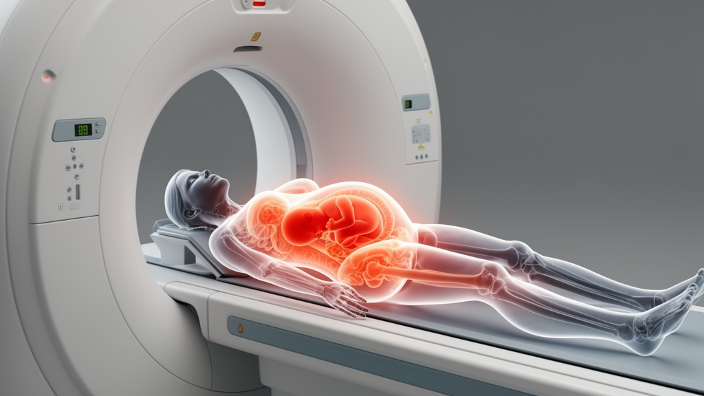
A single scan told two stories at once. In a case report published in the British Journal of Biomedical Sciences, radiologists and obstetric clinicians in Ho Chi Minh City and Vinh Long, Vietnam, describe a 19-year-old woman at 35 weeks’ gestation whose abdominal pain turned out to reflect not one but two simultaneous surgical emergencies: a perforated appendix and acute gallstone cholecystitis. Magnetic resonance imaging, performed without gadolinium contrast and without ionizing radiation, disentangled the overlapping right-sided pathology that ultrasound could only partially explain, allowing surgeons to prioritize the appendiceal process while managing the gallbladder conservatively. The pregnancy then progressed to an uncomplicated term vaginal delivery of a healthy newborn, an outcome the authors attribute to rapid diagnostic clarification and timely operative source control.
The clinical picture was deceptively mild. The woman arrived with a one-day history of dull pain in the right lower quadrant, accompanied by discomfort in the right upper quadrant, the two territories of appendiceal and biliary disease respectively. She reported no fever. On examination, her abdomen was soft, with localized tenderness in the right iliac fossa but no guarding or rebound tenderness. Laboratory testing showed a white blood cell count of 16.34 K/μL with 82.8 percent neutrophils, indicating leukocytosis, and a markedly elevated high-sensitivity C-reactive protein of 239.21 mg/L.
Ultrasound, the standard first-line imaging test in pregnancy, produced a partial answer. It demonstrated a mildly distended gallbladder with a transverse diameter of 39 mm and an approximately 16 mm stone impacted at the gallbladder neck, a finding suggestive of obstructing biliary disease. Fat infiltration in the right iliac fossa was also noted, hinting at inflammation in the appendiceal territory. But the appendix itself could not be confidently visualized, a limitation that becomes more common as gestation advances because the enlarging uterus, bowel gas, and restricted acoustic windows in the right lower quadrant degrade image quality. Fetal assessment showed a viable fetus. With one imaging abnormality identified, a second suspected but unconfirmed, and severe inflammation unaccounted for, the team escalated to abdominal MRI.
The MRI examination relied on axial and coronal T2-weighted sequences, with no gadolinium-enhanced images used for the diagnostic findings. It revealed a markedly enlarged tubular structure consistent with the appendix near the right adnexa, measuring approximately 14 mm in diameter, containing luminal fluid and surrounded by periappendiceal inflammatory change. The inflamed appendix was confirmed on paired imaging planes, supporting a diagnosis of acute appendicitis with significant surrounding inflammation. Critically, the scan also showed free intraperitoneal fluid, predominantly in both iliac fossae with a right-sided predominance, together with inflammatory fat stranding centered in the periappendiceal and paracecal region of the right lower quadrant. That combination raised concern for complicated appendicitis with localized peritonitis, meaning the appendix had likely already breached its wall and was leaking into the peritoneal cavity.
The same scan simultaneously characterized the hepatobiliary abnormalities. The gallbladder was mildly distended at approximately 82 by 29 mm, with an impacted neck stone of roughly 15 mm, gallbladder wall thickening and edema of about 4.5 mm, and pericholecystic inflammatory change, features consistent with acute calculous cholecystitis. The authors note that the triad of an obstructing neck stone, wall edema, and pericholecystic inflammation favored a primary biliary process rather than isolated reactive gallbladder edema, although they caution that because perforated appendicitis with localized peritonitis was present concurrently, a secondary reactive inflammatory component could not be completely excluded. Mild common bile duct dilatation of 6.2 mm was present without MRI evidence of stones within the duct, and available liver biochemistry did not suggest biliary obstruction, placing the obstructing calculus at the gallbladder neck. Mild grade I bilateral hydronephrosis was also noted.
Emergency laparoscopy was performed the following day after multidisciplinary surgical and obstetric evaluation. Entry and port placement were adjusted for the enlarged uterus and established under direct vision using a pregnancy-adapted technique. Intraoperatively, the appendix was identified in the right iliac fossa and appeared gangrenous with distal perforation, dense omental adhesions, fibrinous exudates, and localized purulent fluid in the right hemiabdomen, findings concordant with the MRI evidence of an enlarged inflamed appendix, marked periappendiceal inflammatory change, and peritoneal fluid. Laparoscopic appendectomy was completed successfully. Because the gangrenous, leaking appendix with localized purulent peritonitis represented the immediate source requiring urgent control, appendectomy was prioritized. The gallbladder appeared inflamed but was not tense, and in the absence of MRI evidence of choledocholithiasis or biochemical evidence of biliary obstruction, no gallbladder-directed procedure was performed during the index operation. Histopathological examination confirmed complicated acute appendicitis with transmural inflammation, necrosis, and periappendicitis.
The outcome was favorable on both maternal and fetal fronts. Postoperatively, the patient received antibiotic therapy and routine clinical monitoring, and obstetric ultrasonography confirmed ongoing fetal viability with reassuring parameters. Her recovery was uneventful and she was discharged in stable condition. Subsequent follow-up ultrasonography showed resolution of the gallbladder inflammatory changes, and no additional gallbladder-directed treatment was required. The pregnancy progressed to term, culminating in an uncomplicated vaginal delivery at 38 weeks of gestation. The newborn was healthy, with a birthweight of 3200 grams and Apgar scores of 8 at one minute and 9 at five minutes. The patient expressed relief after her recovery and was reassured by the continued wellbeing of her pregnancy.
Why was this diagnosis so difficult to reach? Acute appendicitis is the most common non-obstetric surgical emergency in pregnancy, yet pregnancy-related physiological and anatomical changes can mask its typical features. As gestational age increases, the enlarging uterus displaces the cecum and appendix superiorly and laterally, shifting the location of maximal tenderness and reducing the reliability of classic peritoneal signs such as guarding and rebound. Nausea, vomiting, mild leukocytosis, and nonspecific abdominal discomfort are common during pregnancy, further reducing the diagnostic specificity of symptoms and basic laboratory tests. Diagnostic delay matters: complicated appendicitis with gangrene, perforation, and peritonitis is associated with substantially worse maternal and fetal outcomes than uncomplicated disease. Yet the competing risk is also real. Large observational data have shown that negative appendectomy in pregnancy is not benign, with fetal loss and adverse obstetric outcomes reported even when the appendix is normal, which is why improved diagnostic accuracy before operative intervention is so important.
MRI occupies a central role in this balancing act because it provides high diagnostic performance without ionizing radiation. Current imaging strategies generally recommend graded compression ultrasound as the first-line test, with MRI as the next step when ultrasound is nondiagnostic. A pregnancy-specific meta-analysis reported MRI sensitivity for appendicitis of approximately 91.8 percent and specificity of approximately 97.9 percent, supporting MRI as a reliable tool to reduce both missed appendicitis and unnecessary surgery. Typical MRI findings supportive of appendicitis include appendiceal dilation, wall thickening, periappendiceal inflammatory change, and free fluid, while signs suggesting perforation include substantial peritoneal fluid, phlegmon, or abscess. For acute cholecystitis, MRI can show gallbladder distension, wall thickening and edema, pericholecystic inflammatory change, and an obstructing calculus. In this patient, MRI simultaneously evaluated the appendix, peritoneal fluid, and biliary system, helping the team identify appendiceal source control as the immediate operative priority while characterizing the gallbladder abnormalities for ongoing surveillance.
The authors frame this report as an illustrative clinical lesson rather than a standardized algorithm. They note that society guidance, including SAGES recommendations, supports laparoscopic approaches for common surgical problems in pregnancy with pregnancy-adapted technique, CO2 insufflation pressures generally in the 10 to 15 mmHg range adjusted to maternal physiology, and multidisciplinary care, while recent SAGES guidelines conditionally recommend laparoscopic cholecystectomy rather than nonoperative treatment for acute cholecystitis in pregnancy. The conservative gallbladder decision in this case should not be interpreted as evidence that acute cholecystitis in pregnancy generally requires nonoperative treatment; the subsequent resolution of gallbladder inflammatory changes without intervention supports the individualized choice here, but clear documentation of biliary findings and postpartum definitive management if symptoms recur remain pragmatic components of care. As a single case report, the study cannot establish comparative effectiveness or a general management pathway, and the published MRI documentation is limited to selected T2-weighted images rather than a complete reproducible acquisition protocol. Some gallbladder wall edema may have been reactive to the concurrent peritonitis. The principal strength is the concordance among clinical assessment, ultrasound, MRI, operative findings, histopathology, and maternal-fetal outcome, demonstrating how cross-sectional imaging can clarify competing right-sided abdominal diagnoses in late pregnancy. The authors conclude that concurrent appendicitis and gallbladder disease should be considered in pregnant patients with mixed right lower quadrant and right upper quadrant pain, and that when ultrasound does not fully explain the presentation in late gestation, MRI can identify concurrent abnormalities and help prioritize timely management without fetal ionizing-radiation exposure.
Subject of Research: MRI diagnosis of concurrent perforated appendicitis and acute calculous cholecystitis in late pregnancy
Article Title: MRI diagnosis of concurrent perforated appendicitis and acute calculous cholecystitis in late pregnancy: a case report
Article References: Huynh, Q. H., Tong Thi, M. K., Nguyen, X. Q., Ho Thi, T. H., & Nguyen, C. P. (2026). MRI diagnosis of concurrent perforated appendicitis and acute calculous cholecystitis in late pregnancy: a case report. British Journal of Biomedical Science, 83, Article 16647. https://doi.org/10.3389/bjbs.2026.16647
Image Credits: AI Generated
Keywords: appendicitis, cholecystitis, MRI, peritonitis, pregnancy, laparoscopy, gallstones, diagnostic imaging, case report, late gestation, emergency surgery, fetal outcome
News Source: Ophelia Keating. (October 26, 2026). MRI reveals two concurrent surgical emergencies in late pregnancy. Scienmag.