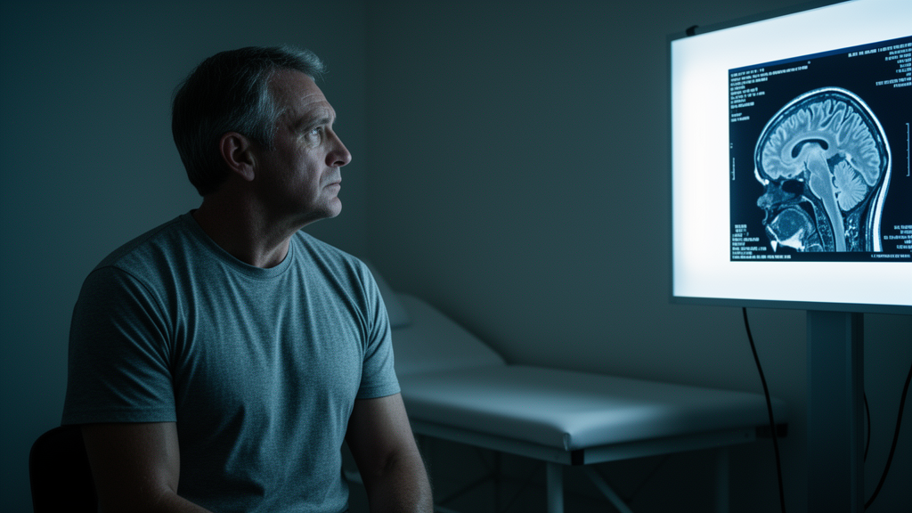
When an athlete takes a blow to the head, the injury that follows is often invisible on the scans that hospitals rely on. Computed tomography and conventional magnetic resonance imaging typically look normal after a mild traumatic brain injury, even as the brain’s functional wiring, blood flow, and chemistry are quietly disrupted. Now, a systematic review from the University of Salzburg has pulled together every multimodal brain imaging study of concussed athletes published to date, and the verdict is sobering: fourteen studies, hundreds of measurements, and still no validated imaging biomarker for concussion. The review, led by Anna E. Bergauer and Fabio Richlan and published in Frontiers in Neurology, systematically searched PubMed and Web of Science up to August 2026 and found that while individual studies report striking alterations in brain connectivity, diffusion metrics, cerebral blood flow, and metabolites, no consistent neuroimaging pattern has emerged across the field.
The stakes could hardly be higher for contact-sport athletes. Concussion risk varies enormously by sport, with rugby carrying the highest rates, followed by American football, ice hockey, and wrestling. Females experience roughly 0.11 more concussions per 10,000 athletic exposures than males, and collegiate athletes are concussed more often than high school players. Most symptoms resolve within 10 to 14 days, but 15 to 30 percent of affected athletes develop persistent post-concussive symptoms lasting beyond three months. Yet physicians currently have no diagnostic, prognostic, or predictive biomarker to guide them. The only blood biomarkers cleared by the Food and Drug Administration, glial fibrillary acidic protein combined with ubiquitin C-terminal hydrolase-L1, serve solely to aid downstream detection of intracranial injury on computed tomography, not to track the subtle injury itself.
Because the brain’s macrostructure usually remains intact after mTBI, diagnosis rests on clinical assessment that blends objective measures, such as balance testing and neurological examination, with symptom self-reports. This strategy carries a real danger: functional recovery often takes longer than symptomatic recovery, so athletes may be medically cleared for return to play while their brains are still compromised. Comorbid conditions such as post-traumatic stress disorder further muddy the picture, since concussion symptoms overlap heavily with psychiatric presentations. The Salzburg reviewers argue that athletes are precisely the population where multimodal imaging could be most informative, because organized sport uniquely permits pre-season baseline assessments, enabling within-subject comparisons that are rarely feasible in the general population, and validated findings could directly protect athletes from premature return-to-play decisions.
To capture the full pathophysiological spectrum, the review focused on studies acquiring two or more complementary imaging modalities within the same participants. The dominant combination was resting-state functional MRI paired with diffusion tensor imaging, used in six studies, followed by fMRI, DTI, and magnetic resonance spectroscopy together in two studies. Arterial spin labeling and MR-phase contrast angiography assessed cerebral blood flow, quantitative susceptibility mapping probed tissue iron contents, and susceptibility weighted imaging searched for microhemorrhage. Each modality contributes a distinct layer of biological information: DTI tracks microstructural integrity of white matter tracts through fractional anisotropy and diffusivity metrics, fMRI maps functional connectivity between brain networks, ASL quantifies perfusion, and MRS measures neurometabolites such as N-acetylaspartate, choline, myo-inositol, and glutamine.
The database search returned 1,302 articles, of which only 14 survived the narrow eligibility criteria requiring athletes with mTBI and genuinely multimodal imaging. Risk of bias, assessed with the Newcastle-Ottawa Scale, was rated low in one study, moderate in twelve, and high in one. The included cohorts skewed young, with five studies examining children under 18 and nine studying young adults under 25. All studies included control groups, though in only nine were the controls athletes playing the same sport. Notably, ten studies assessed concussion history, but just five controlled for it statistically, and no study assessed sub-concussive head impacts, a gap the reviewers flag as critical for disentangling the effects of the most recent injury from cumulative brain exposure.
Grouping findings by post-injury phase, the review found the clearest replication in the transition phase of one to seven days, where two studies independently reported reduced fractional anisotropy and increased mean diffusivity in the corona radiata and internal capsule, the most convergent white matter finding in the entire review. In the acute phase within 72 hours, evidence rested on only five studies, including one reporting a significant cerebral blood flow reduction from 48 to 38 mL/100 g per minute and another documenting a 52 percent drop in glutamine relative to baseline. The subacute phase, spanning three days to three months, drew twelve studies and showed the greatest heterogeneity, with some studies reporting increased fractional anisotropy in the superior longitudinal fasciculus and corpus callosum while others found no longitudinal changes at all.
Functional connectivity findings proved stubbornly inconsistent in direction and region. One study observed hyperconnectivity in posterior brain regions alongside hypoconnectivity anteriorly, another documented a 51.8 percent decline in default mode network connectivity from day one to day seven, and yet another found elevated connectivity within the default mode network during the same window. In the chronic phase beyond three months, only three studies reported significant results, including decreased cerebral blood flow and increased mean diffusivity in large white matter tracts. The single most consistent finding across all four phases was a null result: changes in N-acetylaspartate, a marker of neuronal integrity, never reached statistical significance at any time point in any study that measured it.
Only two longitudinal studies incorporated a neuroimaging baseline, and their samples overlapped, both drawing on female varsity athletes. That scarcity matters enormously. The baseline-enabled study by Schranz and colleagues captured a 52 percent glutamine reduction in the acute phase precisely because each concussed athlete could be compared against their own pre-season scan, a subtle change that would likely have been swallowed by inter-individual variability without the baseline. Manning and colleagues used linked independent component analysis, a sophisticated data fusion technique identifying components with shared variance across modalities, to isolate one component representing the brain’s acute response to concussion, which recovered after three months, and a second representing long-term alterations in corpus callosum diffusion covarying with network connectivity.
Correlations between clinical measures and imaging findings were less consistent than the clinical measures themselves. Symptom scores on the Sport Concussion Assessment Tool and ImPACT reliably peaked at concussion assessment and returned to baseline or control levels at follow-up, yet four studies linked initial symptom severity to neuroimaging recovery trajectories while others found no association between symptom scores and connectivity or blood flow. The reviewers note that the SCAT has limited diagnostic utility beyond three to five days post-concussion, and they suggest broader screening instruments, including the Sport Concussion Office Assessment Tool 6, which assesses vestibular, oculomotor, sleep, headache, cervical, and psychological symptoms, to disentangle concussion-related from co-occurring psychiatric presentations.
The reviewers conclude that current evidence should be regarded as preliminary and hypothesis-generating rather than an established clinical tool. Only three studies conducted prespecified, hypothesis-driven multimodal integration with multiple-comparison correction, while the rest correlated findings that were already significant, providing evidence of statistical covariation rather than mechanistic relationships. No study combined MRI-based with electrophysiological methods, leaving magnetoencephalography and EEG entirely unexplored in this literature despite their complementary temporal precision. The path forward, the authors argue, lies in standardized imaging protocols specifying scanner hardware, sequence parameters, and processing pipelines, adequately powered prospective longitudinal designs with baseline assessments and long-term follow-up, and multimodal data fusion analyses that jointly exploit the complementary strengths of fMRI, DTI, and MEG. Until then, the subtle scars concussion leaves on the athlete brain remain scientifically visible but clinically unusable.
Subject of Research: Neuroscience
Article Title: Multimodal brain imaging of mild traumatic brain injuries in athletes: a systematic review with narrative synthesis
Article References: Bergauer, A. E., & Richlan, F. (2026). Multimodal brain imaging of mild traumatic brain injuries in athletes: a systematic review with narrative synthesis. Frontiers in Neurology, 17, Article 1929474. https://doi.org/10.3389/fneur.2026.1929474
Image Credits: AI Generated
DOI: 10.3389/fneur.2026.1929474
Keywords: concussion, mild traumatic brain injury, multimodal neuroimaging, athletes, diffusion tensor imaging, functional MRI, systematic review, return to play, Multimodal, brain, imaging, mild
News Source: Cassandra Pierce. (October 25, 2026). Concussion Scars the Athlete Brain in Ways Standard Scans Cannot See, Review Finds. Scienmag.