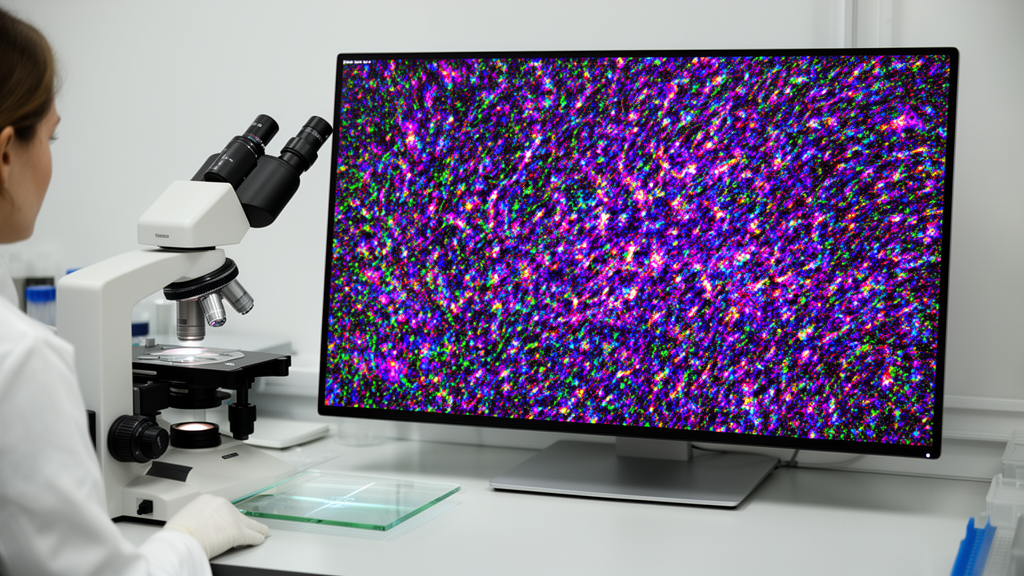
Microscope images of organisms counted in a glass chamber often arrive with a hidden complication: the chamber’s etched measurement grid sits in the focal plane, so its dark lines are recorded across every overlapping image tile. SlidePrep, a new open-source software pipeline, detects those reticle lines, reconstructs the biological content they obscure, registers the corrected tiles and assembles them into a calibrated whole-slide image. The tool can be run from a command line or through a containerized web interface, offering laboratories a way to preserve the acquired specimen across a stitched mosaic while reducing interference from the counting grid.
The problem is especially apparent when a complete chamber is digitized. A Sedgwick-Rafter counting chamber, for example, contains suspended microorganisms observed through an etched reticle that divides the sample volume. Capturing the full chamber can require hundreds of overlapping tiles, each carrying portions of the same high-contrast grid. Although stitching can align those tiles reliably, conventional image processing cannot simply subtract the reticle like additive noise. The etched lines attenuate or block transmitted light, meaning that removing their brightness leaves missing information beneath them rather than restoring the affected specimen.
SlidePrep addresses this structural occlusion with a sequence of image-analysis and inpainting steps. Its core implementation uses Python 3.12 and relies on OpenCV for thresholding, template matching and contour analysis, scikit-learn for a random-forest classifier, LaMa for inpainting and ASHLAR for phase-correlation registration and OME-TIFF assembly. An Angular 21 frontend provides the browser interface, while FastAPI, Celery, Redis, nginx and Docker Compose support asynchronous processing in the web deployment. The current release, version 1.0.4, is distributed under the MIT License.
Each tile is treated independently before the images are joined. First, a three-class multi-level Otsu threshold isolates the darkest intensity population, which corresponds to the etched reticle in brightfield images of the chamber floor, larvae and debris. Connected components are then filtered by shape because cells and pigment deposits tend to be compact, whereas grid lines are elongated. The resulting candidates are located by template matching with a blurred bar representing the expected line thickness and inclination. Lines crossing the full field produce strong detections, whereas fragments at tile edges are weaker and may resemble clipped tissue, so a classifier evaluates their geometric features and retains at most one candidate for each orientation.
After detection, SlidePrep converts the surviving line geometry into a mask. The mask uses rotated minimum-area rectangles, allowing it to follow the reticle’s actual angle rather than assume alignment with the image axes. Each rectangle is widened by a configurable margin so that the inpainting model receives contextual pixels around the obstruction. LaMa then fills the masked area using the unmasked portion of the original tile. The method can continue the outline of an organism crossed by a grid line, although the source evaluation shows that fine internal texture may be lost and that an object boundary cannot always be recovered when no matching continuation is visible on the opposite side of the line.
The corrected tiles are then passed to ASHLAR, which uses the declared acquisition order and overlap only as an initial estimate. Phase correlation with neighboring tiles determines their final positions, while a configurable limit prevents a tile from being displaced beyond a plausible correction. SlidePrep supplies the physical pixel size specified for the acquisition and checks that this calibration is present in the assembled OME-XML, allowing the resulting image to be measured in micrometers in tools such as QuPath and Fiji. A binary mask records every synthesized pixel in the mosaic, enabling downstream analyses to distinguish reconstructed regions from original observations.
Several imaging channels can be processed together, but SlidePrep applies reticle removal asymmetrically. In transmitted-light brightfield, the grid is visible across almost the entire field, whereas in epifluorescence it is apparent mainly where it blocks light from an emitting specimen. The configured brightfield channel is therefore detected and reconstructed, while other channels are carried through without synthesizing intensity. The processed brightfield images also provide the alignment reference for fluorescence, whose background may contain too little signal for dependable registration. A passthrough option allows datasets without a reticle to undergo format conversion and registration without artifact removal.
The web deployment is designed for mosaics that would be impractical to load directly into a browser. For the mussel-larva example, the assembled two-channel image measured 17,640 by 44,447 pixels and occupied 766 MB on disk. SlidePrep converts it into a multiresolution Deep Zoom pyramid with libvips, allowing an OpenSeadragon viewer to load progressively as users pan and zoom from the full chamber to an individual larva. The viewer reports relative zoom rather than objective magnification, and the pyramid does not retain the OME-TIFF’s physical calibration, so the scientifically calibrated file remains available separately for download. A REST API also allows automated workflows to submit jobs, monitor progress, export the mosaic and delete completed datasets without using the frontend.
Performance measurements from the 1,372-tile mussel-larva dataset demonstrate the computational cost of the reconstruction chain. The dataset covered 686 positions in a 14-by-49 raster, with brightfield and epifluorescence images at each position, and was processed on a workstation with an NVIDIA RTX 3050. Across three runs, removing the reticle from the 686 brightfield tiles took a median of 4,698 seconds on the processor and 839 seconds with the graphics processor, corresponding to a 5.6-fold acceleration. LaMa inpainting accounted for 98.4% of the summed per-tile time on the processor and 86.2% on the graphics processor, showing that content reconstruction rather than grid detection dominated the workflow.
The reported detection evaluation, described in a companion paper, produced a tile-level F1 score of 0.9934 and a pixel-level F1 score of 0.9642 across 686 annotated tiles, compared with 0.2974 for an Otsu-binarization baseline using contour detection. These figures describe artifact detection rather than counting accuracy or the fidelity of reconstructed specimen detail. Visual inspection of the assembled mussel-larva mosaic found no obvious periodic pattern at display reductions of about threefold, although a faint pattern became visible at stronger reductions. The authors identified possible causes in residual intensity differences within reconstructed bands and slight illumination differences among tiles, neither of which SlidePrep corrects.
SlidePrep is not yet a universal replacement for every chamber or microscopy workflow. Its classifier is trained on the appearance of a particular reticle, so adapting it to another chamber requires collecting labeled candidates and retraining the model; cross-chamber validation remains future work. The software also assumes that tile filenames follow a documented convention, even though it does not require vendor-specific stage metadata. It corrects structural occlusion rather than photometric non-uniformity, cannot reconstruct a mosaic that has already been assembled, and depends on informative overlap content for accurate registration. The authors plan to validate additional chamber geometries, reduce residual brightness steps in reconstructed bands, quantify reconstruction fidelity against frequency-domain grid erasure, assess effects on downstream counting and add enumeration and morphometry to the viewer.
Subject of Research: Software for removing reticle artifacts, reconstructing tiles and assembling calibrated whole-slide microscopy images.
Article Title: SlidePrep: artifact removal, reconstruction, and visualization of whole-slide images from tiled microscopy acquisitions
Article References: Rodriguez-Conde, I., Campos, C., & Fdez-Riverola, F. (2026). SlidePrep: artifact removal, reconstruction, and visualization of whole-slide images from tiled microscopy acquisitions. SoftwareX, 36, Article 103161. https://doi.org/10.1016/j.softx.2026.103161
Image Credits: AI Generated
DOI: 10.1016/j.softx.2026.103161
Keywords: SlidePrep, whole-slide imaging, microscopy, image inpainting, reticle removal, image stitching, counting chambers, open-source software, mussel larvae, artifact, removal, reconstruction
News Source: Denise Maddox. (October 22, 2026). SlidePrep removes chamber grids and reconstructs whole-slide microscopy images. Scienmag.