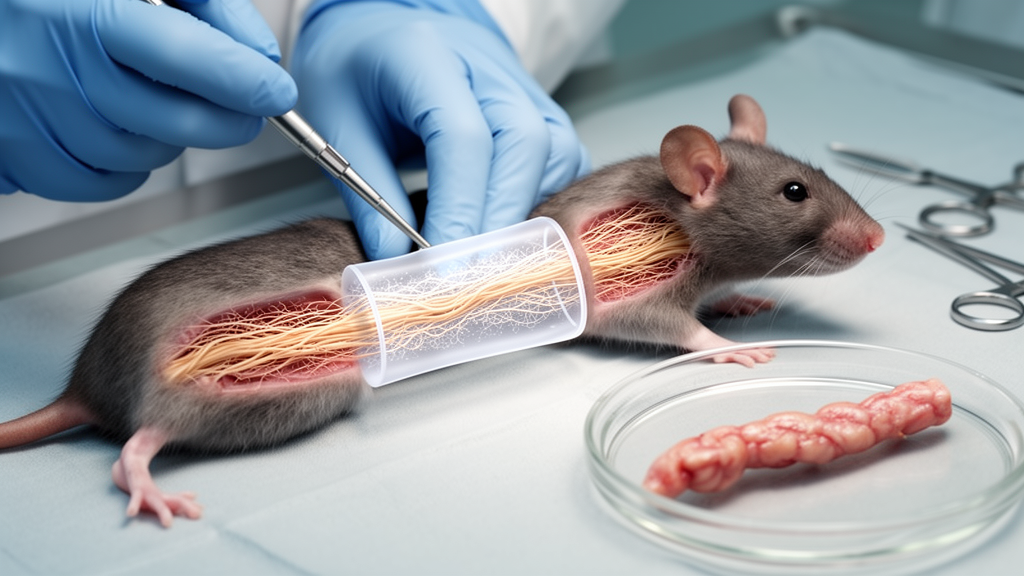
A three-layer artificial conduit containing living Schwann cells restored sciatic nerve connections in rats with performance comparable to a natural nerve autograft, according to a peer-reviewed study in the International Journal of Extreme Manufacturing. The device combined a tough outer support, a porous barrier membrane and an inner set of aligned cell-laden microfilaments designed to guide regenerating axons across a 10 mm nerve gap. Over 16 weeks, the conduits carried electrical signals, supported myelinated nerve bundles and reduced muscle wasting, suggesting that the living structure could function as a tissue-engineered bridge for peripheral nerve repair.
Peripheral nerves normally carry communications between the brain and the limbs, but a severe injury can completely interrupt those pathways. Without direct structural guidance, regenerating nerve fibers may lose their direction, leaving downstream muscles without electrical stimulation and vulnerable to permanent atrophy. Surgeons commonly bridge a damaged nerve segment with an autograft, meaning a healthy sensory nerve taken from elsewhere in the same patient.
Although this approach is clinically established, it requires sacrificing another nerve and can leave donor-site numbness, chronic pain or a reduced supply of usable tissue. Hollow plastic conduits avoid that surgical cost, but the release notes explain that wider gaps can be difficult to cross because the tubes may not reproduce the physical cues and biochemical support needed by growing axons. The team addressed that problem by integrating three fabrication techniques on one direct-writing platform: melt writing for the structural outer layer, electrospinning for the porous middle membrane and low-temperature cell patterning for the innermost living tracks.
The conduit was built continuously rather than transferring fragile components between separate machines. Its outer layer consists of an open polycaprolactone mesh that functions as an exoskeleton and resisted compression and suturing forces of up to 1.64 N. The middle layer is a nanofiber membrane with pores averaging 0.55 micrometers across. These openings permit the movement of oxygen and nutrients while discouraging scar-forming connective tissue from entering the conduit.
The innermost layer contains Schwann-cell-bearing microfilaments, creating a biological trackway for nerve regrowth. At the core of the work was a bioprinting challenge involving the printing of living Schwann cells, the supporting cells that normally help insulate and direct peripheral nerve fibers. Electro-assisted bioprinting can pull cell-containing ink through fine nozzles into microfibers, but high-resolution printing creates a damaging trade-off.
Viscous inks tend to retain their deposited shape, yet forcing them through narrow nozzles produces strong shear stress that can rupture cell membranes. Diluting the ink reduces that stress, although the resulting filaments can collapse into puddles after deposition. Chemical hardeners may be toxic to the cells, while the relatively high voltages used to form fine fibers can also damage living material.
The researchers managed this conflict with temperature. Their bioink contained alginate, polyethylene oxide and thermosensitive gelatin methacryloyl, together with living Schwann cells. The nozzle was held at 25 °C, where the mixture was fluid enough to pass through the nozzle without imposing the shear associated with a thicker formulation.
A chilled collection plate at 4 °C rapidly stabilized each deposited jet before it could spread. Under a comparatively gentle electric field of 1.0 kilovolt, the system produced continuous microfilaments measuring about 36 micrometers in diameter. Temperature therefore served as a way to control the material’s flow during printing and its stability immediately after deposition.
In laboratory cultures, nerve cells attached to the printed tracks organized into dense, directionally aligned ribbons rather than dispersing randomly. RNA sequencing suggested a molecular explanation for this behavior. Contact with the microfilaments, together with chemical signals supplied by the Schwann cells, activated axon-guidance genes including Ephb1 and Gnai1.
Those signals were associated with cytoskeletal programs that steer elongating axons. At the same time, expression linked to programmed cell death and routine cell proliferation decreased, while the cells redirected their activity toward extending elongated signaling structures. These observations concern laboratory cultures and gene-expression patterns; the release does not report a separate clinical assessment of the underlying molecular mechanisms.
When implanted across 10 mm gaps in rat sciatic nerves, the composite conduits were evaluated over 16 weeks against natural nerve autografts and conduits without living cells. Regenerating nerve bundles in the living devices developed myelin insulation averaging 1.02 micrometers in thickness, close to the 1.15-micrometer average measured in the autografts. Repaired nerves in the living-conduit group conducted impulses at 5.67 meters per second, compared with 5.88 meters per second for the autograft control.
The living-conduit rate was nearly double the rate measured in the acellular conduits. These findings indicate comparable functional performance between the two repair strategies in this rat model, rather than proof of equivalence in people. The experiment also shows that the living patterned tracks supported a different outcome from empty conduits, although the release does not provide a numerical conduction rate for the acellular group.
Functional recovery extended beyond the measured conduction velocity. Severed connections normally leave distal calf muscles vulnerable to denervation atrophy. The living conduits preserved approximately 49% of distal calf muscle wet weight, whereas empty conduits preserved less than 24%.
Walking trials also showed restoration of normal hindpaw coordination in animals receiving the living conduits. Together, the electrophysiological and behavioral results suggest that the engineered bridge did more than provide a passive path: the patterned Schwann-cell tracks supported organized nerve regrowth strong enough to reduce downstream muscle loss and recover coordinated movement during the study period. The reported results are therefore not limited to an electrical measurement; they include anatomical evidence of myelinated bundles and functional measures of muscle preservation and movement.
The study offers a manufacturing proof of concept for multilayered implants containing living cells, but the evidence remains preclinical. Before human testing, the researchers will need to test the conduits across gaps measured in multiple centimeters in large animal models. The team must also establish low-temperature sterilization methods that do not kill the embedded Schwann cells and adjust the degradation rate of the polycaprolactone shell so that it disappears at a pace suited to the slower timetable of human nerve healing.
The reported results support further large-animal validation, not an immediate clinical recommendation. If that next phase succeeds, the approach could offer an alternative for patients who cannot donate another healthy nerve or do not have enough tissue for a conventional autograft. For patients, the significance of the work is that it combines several functions in one device: mechanical support, selective exchange with the surrounding environment and a living, aligned substrate for nerve fibers.
The conduit is not simply a hollow tube, because its inner tracks are populated with cells that can provide biochemical cues while the printed architecture supplies physical direction. At the same time, the outer polycaprolactone layer is designed to withstand handling while the middle membrane limits inward connective-tissue growth. The result is a compact example of how melt writing, electrospinning and low-temperature bioprinting can be combined without moving delicate constructs between separate machines.
The study’s central claim is a preclinical one, supported in rats: under the reported conditions, a living bioprinted conduit supported nerve regeneration over a 10 mm gap with functional outcomes close to those of a natural autograft. Its broader usefulness will depend on validation across longer gaps, sterilization methods compatible with living cells, shell degradation matched to healing time, and eventual testing in humans.
Subject of Research: Preclinical bioprinted nerve conduits for peripheral nerve regeneration in rats
Article Title: Living "railway tracks" bioprinted inside artificial conduits match gold-standard nerve repair
Article References: Living "railway tracks" bioprinted inside artificial conduits match gold-standard nerve repair. Original publication (Bibliographic details unavailable.)
Image Credits: AI Generated
DOI: Not provided
Keywords: peripheral nerve repair, 3D bioprinting, Schwann cells, nerve guidance, tissue engineering, sciatic nerve, rat study, aligned microfilaments, biofabrication, electrospinning, Living, railway
News Source: Denise Maddox. (October 22, 2026). Bioprinted living nerve conduits match autografts in rat study. Scienmag.