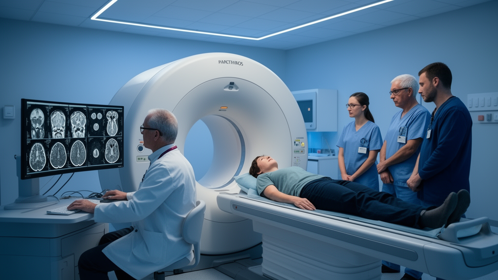
An exploratory MRI study of 13 people with brain tumors involving the supplementary motor area found structural and functional network differences before surgery. Diffusion-tensor imaging showed signs of white-matter disruption in pathways associated with the tumor, while resting-state functional MRI revealed both reduced and increased coupling across sensorimotor and cognitive networks. Because the cohort was small, heterogeneous and lacked systematic neuropsychological testing, the findings cannot show that the imaging changes caused specific cognitive or motor impairments or reliably predict recovery after tumor removal.
The supplementary motor area, or SMA, participates in initiating voluntary movement and has connections with regions involved in language and cognitive control. Patients whose tumors involve this area may develop “SMA syndrome” after surgery, a condition that can include temporary inability to move the opposite side of the body and, when the dominant hemisphere is affected, mutism or expressive speech impairment. Symptoms commonly improve over subsequent months, although motor recovery can remain incomplete. Researchers at RWTH Aachen University investigated whether abnormalities in brain connectivity were already present before treatment, when most patients in this study retained substantial motor and speech function.
The investigators compared 13 SMA-tumor patients with 13 age-matched healthy controls. The participants underwent preoperative diffusion-tensor imaging and resting-state functional MRI on a 3 Tesla scanner. The patient group had a mean age of 49 years and included several tumor types, including gliomas, metastases and a meningioma. Only two patients had minor motor deficits and one had minor speech impairment before surgery. For group-level analyses, images from patients with right-sided tumors were flipped so that lesioned and contralesional hemispheres could be compared consistently, with an equivalent number of control images also flipped.
Structural analysis focused on the frontal aslant tract, which connects the SMA with the ipsilateral inferior frontal gyrus, and on transcallosal pathways linking the two SMAs and each SMA with primary motor cortex in the opposite hemisphere. In the frontal aslant tract, patients showed higher mean diffusivity and axial diffusivity on the tumor side than on the contralateral side, a pattern interpreted by the researchers as microstructural disintegration. The lateralization effect for fractional anisotropy was close to the significance threshold, while radial diffusivity showed a similar trend. These differences suggest that structural changes around the tumor may extend beyond the lesion’s macroscopically visible boundaries.
Connections between the ipsilesional SMA and the contralesional primary motor cortex also appeared altered. Their normalized tract volumes were significantly lower in patients than in controls, with a reported group p-value of 0.005, independent of hemisphere. Fiber length differed between hemispheres in patients: the contralesional SMA-to-ipsilesional motor-cortex pathway was longer than the pathway running in the opposite direction, whereas no comparable difference was detected in controls. Axial diffusivity also differed between groups, while mean diffusivity and radial diffusivity were reported as trends. Together, these observations point to disruption and asymmetry in an interhemispheric motor pathway before surgery, although individual diffusion measures do not directly establish how the pathways would function after resection.
Structural connectivity between the homologous left and right SMAs did not show the same pattern. Tract volume was not significantly reduced in the patient group, and some patients appeared to have more normalized SMA-to-SMA fibers than controls, but the wider variation prevented a significant group difference. In an exploratory post hoc comparison, patients without residual motor deficits at follow-up tended to have greater SMA-to-SMA tract volume than those with incomplete motor recovery; the difference was not statistically significant. The result cannot establish that larger or better-preserved SMA-to-SMA connections caused better recovery. It instead identifies a possible anatomical feature for evaluation in adequately powered, longitudinal studies.
Resting-state functional MRI produced a mixed pattern across large-scale networks. Group independent-component analysis identified 40 network components, of which 14 represented the sensorimotor, default mode, frontoparietal, salience or attention networks and were selected for region-to-region comparisons. Patients had reduced coupling between the lateral default mode and frontomedian networks, between dorsal-attention and salience components, and between the ipsilesional frontoparietal network and the anterior default mode network. These were associations in preoperative imaging data. Because the researchers did not administer systematic neuropsychological tests, they could not determine whether the lower coupling corresponded to measurable weaknesses in attention, executive function, language or another cognitive process.
Other connections were stronger in the patient group. Coupling increased between the anterior and lateral default mode networks, between the anterior default mode and lateral sensorimotor components, and between medial and lateral sensorimotor components. Connectivity also increased between the frontomedian network, which included the SMA, and the contralesional frontoparietal network. The three clusters showing group differences met a false-discovery-rate-corrected significance threshold of p < 0.046. The authors described the combination of structural disruption, decreased coupling in some networks and increased coupling in others as potentially compatible with both disintegration and reorganization. The MRI observations alone cannot determine whether stronger functional coupling was beneficial compensation, pathological hyperactivity, functional disinhibition, a direct tumor-neural effect or altered neurovascular coupling.
Postoperative records illustrated the clinical course but did not establish prognostic value for individual imaging measurements. Eleven of the 13 patients, or 85%, developed SMA syndrome soon after surgery, with contralateral akinesia; five of eight patients with left-hemispheric tumors also had speech deficits. At a median follow-up of 133 days, motor deficits had improved in nine patients and speech deficits had resolved in all affected patients. Nevertheless, six of the 12 patients with evaluable follow-up data had incomplete motor recovery. The cohort was too small and clinically heterogeneous to determine reliably whether particular baseline connectivity patterns predicted postoperative motor or speech outcomes. Tumor growth behavior, infiltration, other clinical characteristics and antiseizure medication were not fully accounted for in the retrospective analysis.
Technical limitations also constrain interpretation. Deterministic tractography can terminate prematurely or steer fibers incorrectly in regions with crossing pathways, substantial edema, severe tumor distortion or degraded signal, and inter-rater reliability was not assessed. Diffusion acquisition parameters varied for some participants following a scanner software upgrade. Phantom tests indicated stable or improved scanner performance, and identical processing pipelines were used across groups, but protocol-related effects could not be completely separated from patient-group effects. Several exploratory interaction findings did not remain significant after correction for multiple comparisons. The study also did not distinguish the SMA proper from pre-SMA because tumor growth can alter the normal cytoarchitecture that defines their boundaries.
The study therefore offers preliminary evidence that structural and functional connectivity abnormalities can be detected before surgery in people with SMA-region tumors. It does not establish that those abnormalities cause SMA syndrome, reflect specific cognitive deficits or can be used to guide treatment or predict an individual patient’s recovery. Larger, preferably multicenter cohorts with more uniform tumor types, standardized longitudinal imaging and formal neuropsychological assessment are needed. Such follow-up would test whether increased functional coupling before surgery represents helpful compensation or maladaptive reorganization, and whether baseline “connectome” measures can eventually contribute to individualized counseling and postoperative outcome prediction.
Subject of Research: Preoperative structural and functional brain connectivity in patients with supplementary motor area tumors
Article Title: Tumor lesions in the supplementary motor area may induce large-scale sensorimotor and cognitive network alterations— A functional and structural connectivity analysis
Article References: Na, C.-H., Sijben, R., Posnansky, O., Wiesmann, M., Clusmann, H., Delev, D., & Rodriguez-Raecke, R. (2026). Tumor lesions in the supplementary motor area may induce large-scale sensorimotor and cognitive network alterations— A functional and structural connectivity analysis. Frontiers in Neurology, 17, Article 1878278. https://doi.org/10.3389/fneur.2026.1878278
Image Credits: AI Generated
DOI: 10.3389/fneur.2026.1878278
Keywords: supplementary motor area, brain tumors, structural connectivity, functional connectivity, diffusion tensor imaging, resting-state fMRI, SMA syndrome, motor recovery, cognitive networks, Tumor, lesions, supplementary
News Source: Colin Clarke. (October 21, 2026). SMA tumors linked to preoperative brain-network changes in small MRI study. Scienmag.