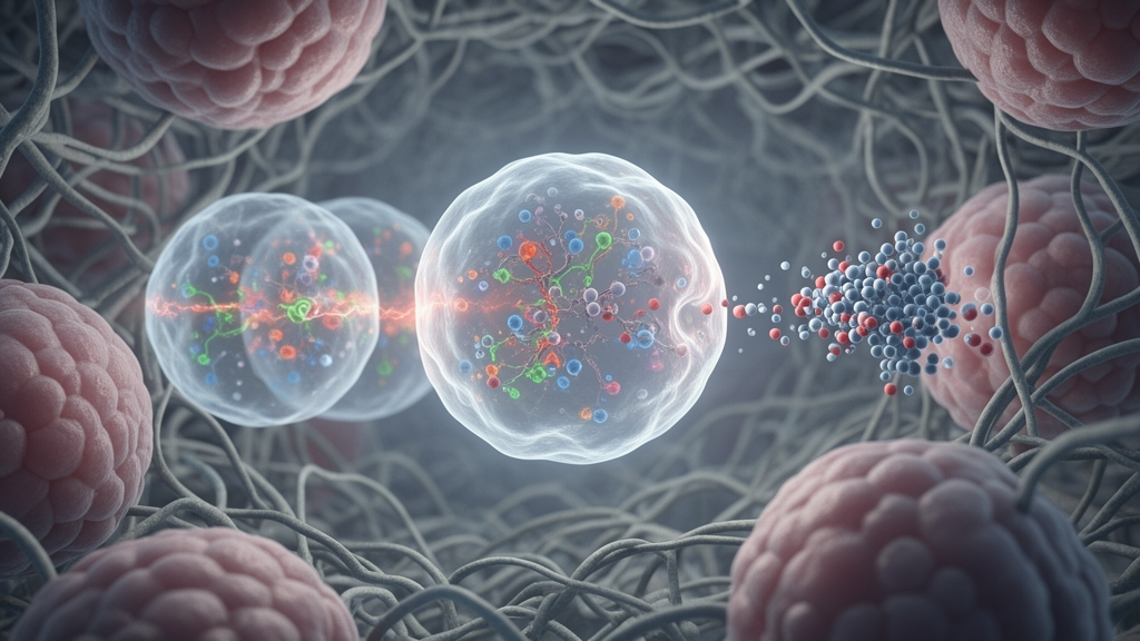
Researchers at the University of California, San Diego, have developed two virtual-cell approaches that use four-dimensional microscopy to represent mitochondrial responses to drugs in human cancer cells. A deep-learning system called MitoSpace, trained on 40,000 movies showing cells treated with 25 compounds, predicted cellular energetic state from mitochondrial shape and movement and distinguished drug-response patterns with 75% accuracy, compared with 56% for a model trained on conventional two-dimensional images. In a separate study, a physics-based digital twin reproduced the reduced movement and altered fusion and fission rates of mitochondrial networks after microtubules were partially broken down by nocodazole. Both peer-reviewed studies, published in Cell, were conducted with cultured cell models and provide computational tools for studying cellular dynamics rather than evidence of clinical efficacy.
Mitochondria are usually illustrated as separate kidney-bean-shaped organelles, but the structures inside living cells form a dynamic, interconnected network. They continually split and fuse and are transported to locations where energy is required. Because mitochondrial networks change shape according to cellular condition, their form and movement can serve as indicators of biological state or responses to treatment. Historically, however, many cell-based screens have used flat, still images. The UC San Diego studies instead employed 4D lattice light-sheet microscopy, which records cellular structures in three dimensions through time and generated the dynamic mitochondrial data used to build both kinds of virtual cell.
For the MitoSpace study, the researchers treated cancer cells with 25 compounds known to perturb mitochondria through different mechanisms. This produced a library of 40,000 single-cell 4D movies. The model was designed to identify patterns without requiring researchers to manually label the images with predefined categories. It learned representations that distinguished one cell from another and, after training, arranged cells into groups reflecting similar responses. The work tested whether information about a cell’s energetic condition could be recovered from the shape and movement of its mitochondria alone, across the dataset’s 26 drug conditions.
MitoSpace predicted cellular energetic state without being given that answer during training, supporting a strong relationship between mitochondrial form, movement and cell function in the tested system. The model’s performance also improved when the input data preserved four-dimensional structure. It distinguished among drugs with 75% accuracy when trained on the 4D movies, whereas the version trained on the flat 2D images commonly used in large-scale drug screens achieved 56% accuracy. This comparison indicates that temporal and three-dimensional information helped the system identify response patterns associated with different mechanisms of mitochondrial perturbation, although the study does not establish that every compound or cellular state will be classified equally well.
The researchers also tested whether the representation learned from the movie library could be applied without retraining. MitoSpace organized examples of drugs it had not previously seen and sorted human lung organoid cells according to developmental stage. These findings show that the model could generalize beyond the exact drug examples used to train it in the experiments reported. They do not demonstrate that it can predict clinical outcomes, and the evidence comes from selected laboratory cell systems rather than human trials. Even so, the results suggest that 4D mitochondrial recordings may provide information that could support analysis of large screening datasets without relying only on the conventional static-image approach.
The second study took a physics-based rather than purely machine-learning approach. Using specialized image-analysis software, the researchers mapped mitochondrial locations and the microtubule tracks along which the organelles travel in a living cancer cell. They also added motor proteins responsible for mitochondrial transport, applying rates established in earlier research. Parameters in the resulting digital twin were adjusted until its mitochondrial behavior matched measurements from the real cell. The simulation incorporated the laws of motion and was constrained by microscopy-derived positions, transport tracks and previously established motor-protein behavior rather than by a statistical model trained solely to classify outcomes.
To evaluate whether the digital twin could reproduce an imposed change, the team asked it to predict the mitochondrial response to nocodazole, a drug that partially breaks down microtubules. Without changing any model parameters, the digital twin generated the reduced mitochondrial motion and fusion and fission rates observed in real cells exposed to the drug. This test indicates that its transport rules captured enough of the measured network behavior to reproduce the response to microtubule disruption. It also enabled a direct side-by-side comparison between a physics-based virtual cell and an actual 3D microscopy movie, an analytical capability the authors described as unprecedented.
The authors propose that digital twins could help investigate potential effects of drugs, disease mutations and cellular-engineering designs before the corresponding laboratory experiments are performed. The approach is especially relevant to Cal27, a head-and-neck cancer cell line in which mitochondrial regulation remains an active research question. The study itself does not identify or validate a cancer treatment, and its demonstrated result is limited to reproducing mitochondrial-network behavior in the tested cellular model. The team has filed for patent protection on the technology, while corresponding author Johannes Schöneberg has started a company based on it.
Schöneberg said the studies reflect a need to capture more than a single snapshot when building computational models of cells. “A cell is a four-dimensional object: it has depth and it never stops moving,” he said. “Virtual cells need to be built on data that captures that fact.” The two approaches address different aspects of the same problem: MitoSpace searches rich datasets for biologically meaningful patterns, whereas the physics-based twin explores the physical processes that can produce those patterns. The researchers plan to combine the methods into one workflow and extend the models beyond mitochondria by incorporating additional organelles.
The longer-term objective is to model groups of cells interacting as tissues and, eventually, to simulate more realistic human biology. Schöneberg said such tissue modeling could eventually inform clinical treatment, but that remains a future goal rather than a result demonstrated in the studies. At present, the evidence consists of cultured-cell experiments, a deep-learning model evaluated across selected compounds and organoid samples, and a digital twin tested against microtubule disruption in one cancer-cell system. Together, the studies show that 4D imaging can supply richer inputs for virtual-cell models and that both learned representations and explicit physical simulations can reproduce selected mitochondrial responses to experimental perturbations.
Subject of Research: 4D microscopy-based virtual cells for modeling mitochondrial drug responses in human cancer cells
Article Title: Virtual cells built from 4D AI models and "Digital twins” could speed up drug discovery
Article References: Virtual cells built from 4D AI models and "Digital twins” could speed up drug discovery. Original publication (Bibliographic details unavailable.)
Image Credits: AI Generated
DOI: Not provided
Keywords: virtual cells, 4D microscopy, mitochondrial dynamics, deep learning, digital twins, drug screening, cancer cells, mitochondria, Cell journal, UC San Diego, Virtual, cells
News Source: Nathaniel Bowman. (October 21, 2026). 4D virtual cells model drug responses in human cancer cells. Scienmag.