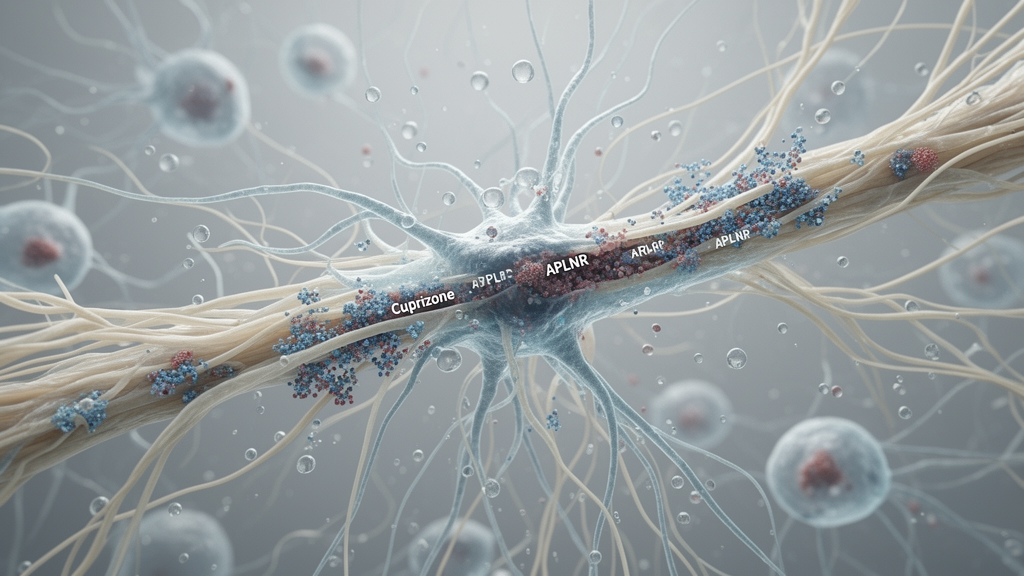
The apelin receptor, APLNR, became strongly associated with markers of reactive astrocytes in the corpus callosum of male mice during cuprizone-induced demyelination, according to an experimental study in Scientific Reports. The association varied by brain region and treatment phase: overlap between APLNR and the astrocyte markers GFAP and CD44 was greatest during active demyelination, particularly in the lateral corpus callosum. These findings establish a spatial and temporal association, not a causal effect of APLNR on myelin repair.
Researchers assigned 30 eight-week-old male C57BL/6 mice to control, demyelination and remyelination groups, with 10 animals per group. Demyelination was induced by administering drinking water containing 0.2% cuprizone for five weeks, followed by a five-week recovery period after cuprizone withdrawal. Investigators used immunohistochemistry, immunofluorescence and confocal microscopy to examine the lateral and medial corpus callosum for myelin basic protein, or MBP; GST-π-positive mature oligodendrocytes; GFAP-positive reactive astrocytes; CD44-positive glial populations; and APLNR.
Cuprizone caused marked myelin loss and depletion of GST-π-positive mature oligodendrocytes. These changes were followed by partial recovery of MBP and the mature oligodendrocyte population during the recovery phase. The regional response was uneven: MBP loss was greater in the lateral corpus callosum than in the medial region, matching the established vulnerability of the rostral corpus callosum in this model. GFAP-positive astrocytic reactivity increased markedly during demyelination and remained more persistent in the lateral region during recovery. CD44 showed a distinct distribution that depended on both experimental phase and location.
APLNR immunoreactivity was minimal in control animals, rose markedly during demyelination and remained elevated during remyelination. Its spatial overlap with GFAP and CD44 was strongest during active demyelination, especially in the lateral corpus callosum, while correlation analyses indicated relationships between APLNR and the astrocytic markers that varied by region and phase. The study therefore identifies APLNR as part of an organized glial response to experimentally induced demyelination. It does not demonstrate that APLNR drives astrocyte remodeling, promotes oligodendrocyte recovery or improves neurological function, and the authors state that the functional significance of the association remains unresolved.
Subject of Research: Experimental mouse study of APLNR-associated astrocytic responses during cuprizone-induced demyelination and recovery
Article Title: Dynamic association of APLNR with astrocytic remodeling in the cuprizone-treated corpus callosum
Article References: Gaydarski, L., Petrova, K., Stamenov, N., Iliev, A., Stanchev, S., Rashev, P., Pupaki, D., Mourdjeva, M., Kostadinova, I., & Landzhov, B. (2026). Dynamic association of APLNR with astrocytic remodeling in the cuprizone-treated corpus callosum. Scientific Reports. https://doi.org/10.1038/s41598-026-76121-8
Image Credits: AI Generated
DOI: 10.1038/s41598-026-76121-8
Keywords: APLNR, apelin receptor, astrocytes, cuprizone model, demyelination, remyelination, corpus callosum, glial response, myelin, C57BL/6 mice, Dynamic, association
News Source: Denise Maddox. (October 21, 2026). Dynamic association of APLNR with astrocytic remodeling in the cuprizone-treated corpus callosum. Scienmag.