In an in vitro study using mesenchymal-like SK-Hep1 cells, interleukin-1β (IL-1β) enhanced several effects induced by transforming growth factor-β1 (TGF-β1), including fibronectin and N-cadherin expression, cell migration and cell invasion. The reported abstract further indicates that IL-1β increased phosphorylation of p38 mitogen-activated protein kinase (MAPK), while also enhancing TGF-β1-associated SMAD2/3 phosphorylation and movement of SMAD2/3 into the nucleus. The findings identify an association between p38 MAPK activity and TGF-β/SMAD signaling in this experimental system.
Researchers treated SK-Hep1 cells with TGF-β1, IL-1β or the two agents together. Western blotting and immunofluorescence were used to assess mesenchymal marker expression, while wound-healing and Matrigel transwell invasion assays evaluated migration and invasion. The selective p38 inhibitor SB202190 was then used to examine whether the observed cellular responses depended on p38 signaling. These methods confined the investigation to cultured cells and did not involve animals, patients or clinical outcomes.
According to the abstract, TGF-β1 induced fibronectin and N-cadherin expression and increased the cells’ migratory and invasive capacities. Adding IL-1β further enhanced those responses. At the signaling level, IL-1β markedly increased p38 MAPK phosphorylation and strengthened TGF-β1-mediated SMAD2/3 phosphorylation and nuclear trans-localization. SB202190 decreased fibronectin and N-cadherin expression and reduced migration and invasion, supporting a functional contribution from p38 MAPK to the measured phenotype.
The study therefore supports the conclusion that IL-1β can potentiate selected TGF-β1-induced responses in SK-Hep1 cells through a p38 MAPK pathway linked to TGF-β/SMAD signaling. However, the evidence presented here comes from a single cell line and its reported in vitro assays. The summary does not establish whether the same mechanism operates in primary tumor tissue, other hepatocellular carcinoma models, immune cells or patients, and it provides no evidence that p38 inhibition or anti-inflammatory treatment improves clinical outcomes. The authors’ proposal for further investigation of anti-inflammatory strategies should consequently be understood as a research direction rather than a demonstrated therapeutic approach.
Subject of Research: In vitro analysis of IL-1β modulation of TGF-β1-driven mesenchymal phenotypes and migration in SK-Hep1 cells
Article Title: Interleukin-1β potentiates transforming growth factor-β-induced fibronectin expression, migration, and invasiveness of mesenchymal-like SK-Hep1 cells via p38 mitogen-activated protein kinase–SMAD2/3 crosstalk
Article References: Fatima, S., Regmi, B., Hwang, S., Kim, M. Y., Baik, S. K., Jung, P. Y., & Eom, Y. W. (2026). Interleukin-1β potentiates transforming growth factor-β-induced fibronectin expression, migration, and invasiveness of mesenchymal-like SK-Hep1 cells via p38 mitogen-activated protein kinase–SMAD2/3 crosstalk. Molecular Biology Reports, 53(1), Article 1580. https://doi.org/10.1007/s11033-026-12763-1
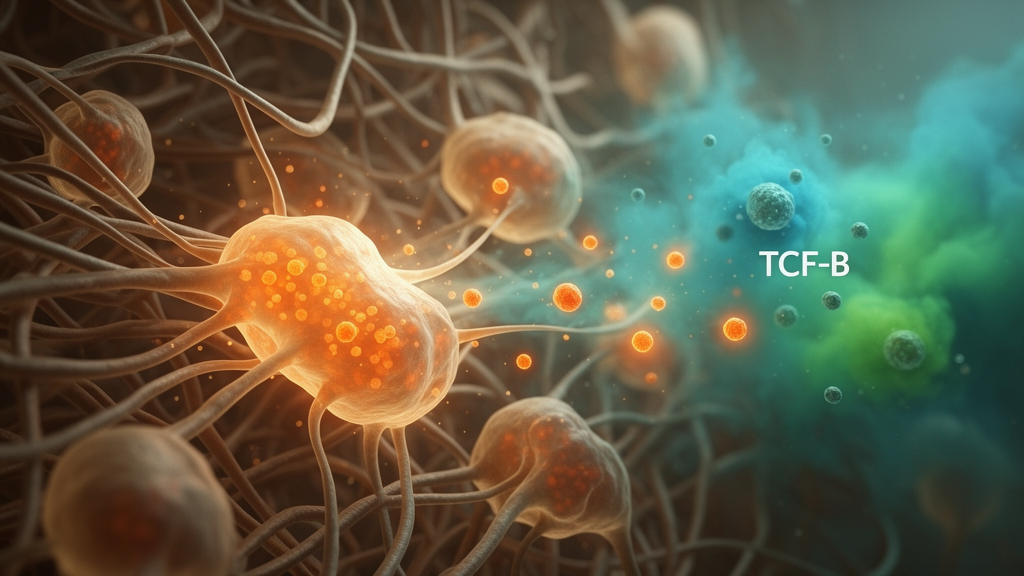
Image Credits: AI Generated
DOI: 10.1007/s11033-026-12763-1
Keywords: IL-1β, TGF-β1, SK-Hep1 cells, hepatocellular carcinoma, p38 MAPK, SMAD2/3, fibronectin, N-cadherin, cell migration, cell invasion, mesenchymal phenotype, Interleukin-1
News Source: Nathaniel Bowman. (October 20, 2026). Cell study finds IL-1β amplifies TGF-β1 effects in SK-Hep1 cells through p38 MAPK-linked SMAD2/3 signaling. Scienmag.