A 2026 review concludes that combining artificial intelligence with four-dimensional bioprinting could improve the design of implants that change shape after implantation, but emphasizes that the field remains dominated by laboratory, animal and simulation evidence rather than successful clinical reconstruction. The authors examine potential applications across craniofacial bone defects, facial skin and mucosa, peripheral nerves and vascularized composite tissues. Their central finding is that AI may help overcome three recurring bottlenecks: selecting materials with the required properties, controlling printing processes, and predicting how a printed structure will deform. However, the review identifies unresolved problems in mechanical strength, reproducibility and access to clinically representative data that prevent the technology from being presented as an established treatment.
The motivation comes from the difficulty of rebuilding the highly intricate structures of the cranium, maxilla, mandible, orbital cavity and nose. Conventional repair can be limited by donor-site injury, the finite amount of available tissue and imperfect anatomical or functional matching. Craniomaxillofacial subunit transplantation, also called facial vascularized composite allotransplantation, transfers several tissue types together, including skin, mucosa, blood vessels, muscle and bone, in an attempt to replace a facial unit as a coordinated whole. This approach is anatomically feasible, but it requires lifelong immunosuppression and therefore carries the risks of rejection and medication toxicity, while also depending on a limited supply of donors. The review presents bioprinting as a possible route toward regenerative reconstruction that could reduce dependence on donor tissue.
Four-dimensional bioprinting extends conventional three-dimensional printing by adding controlled change over time. A printed construct can be compressed for delivery through a small incision and then recover a patient-specific three-dimensional shape after encountering a stimulus such as heat, moisture, altered pH or a magnetic field. This capacity could allow a scaffold to pass through a narrow surgical access route before expanding to match an irregular defect. Shape-memory polymers offer adjustable stiffness and degradation and may be useful for nerve conduits and conformal shells around non-load-bearing areas. Nickel-titanium shape-memory alloys can provide active shape recovery and superelasticity, but their non-degradability and possible nickel-ion release constrain prolonged use. Hydrogels and cell-laden bioinks are much softer, generally occupying the kilopascal range, and are more appropriate for skin, mucosa, nerve and other soft-tissue applications.
AI enters the workflow because the behavior of smart materials depends nonlinearly on composition, geometry, temperature, stress history and deformation rate. Conventional trial-and-error screening becomes especially inefficient when many formulation and structural variables must satisfy conflicting requirements. The review describes support vector regression, extreme gradient boosting, Gaussian process regression, artificial neural networks and physics-informed neural networks as tools for predicting material behavior. In one study, a Gaussian process regression model optimized processing conditions for thermally responsive microneedle patches; in a diabetic rodent model, the resulting patches produced near-complete wound closure within 10 days, with enhanced re-epithelialization and collagen remodeling. Separately, an artificial neural network reduced errors in predicting the ultimate tensile strength and shape-memory activation of a 4D-printed polymer filament by about 40% to 50% relative to linear regression and LASSO methods.
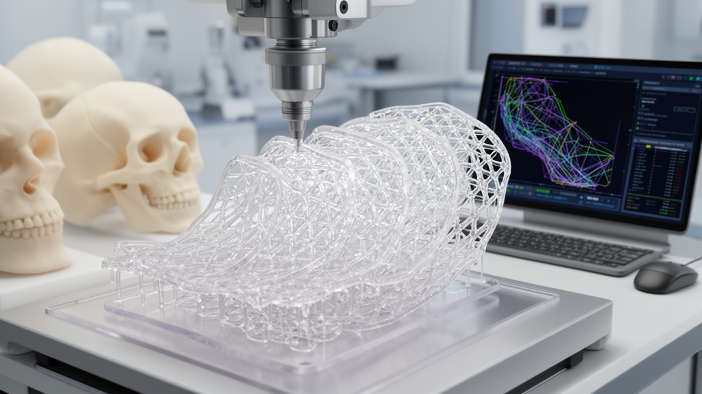
A physics-informed neural network reported in the review required only two temperature-stress curves to predict stress evolution and free-strain responses in amorphous shape-memory polymers. AI can also optimize the manufacturing process itself. A deep reinforcement-learning method tested for laser-melting path planning increased path-generation efficiency by 99.8% while reducing structural deformation by 64.0% and residual stress by 21.8%, according to the review. The authors also identify inverse design as a way to work backward from a desired anatomical outcome. Fully convolutional networks trained on more than 100,000 nonlinear finite-element datasets predicted printable mesh configurations from three-dimensional depth images with an intersection-over-union score above 90%.
Patient-specific facial structures have also been used to test the transition from computational design to physical fabrication. One dual-material system adjusted material ratios and rib-arc angles to create different deformation modes resembling a protruding nasal unit. The reported bioprinted facial masks and thin nasal shells differed from their simulation predictions by less than 10% of their overall dimensions, and transfer learning was used to extend the models from simulated data to authentic human facial anatomy. These results show feasibility within controlled settings, but they do not establish performance after implantation. Most of the models discussed were trained on finite-element simulations, leaving a possible gap between virtual predictions and the behavior of physical materials in living tissue.
Several studies in the review tested shape-adapting scaffolds in rodent bone-defect models. A dual-function scaffold with a tunable transition temperature of 37 to 42 degrees Celsius could be compressed and then triggered by near-infrared light to recover an anatomically matching shape. Hydrogel layers containing bioactive composites were intended to support stem-cell osteogenic differentiation, and the scaffold showed conformity and bone formation in rat cranial defects. Separately, a gradient alginate scaffold developed curvatures from 0.05 to 0.446 per millimeter and accelerated new bone formation in rodent models. Another PLLA scaffold containing magnetic nanoparticles recovered more than 98% of its shape within 40 seconds under external magnetic stimulation. It also suppressed NLRP3 inflammasome activation in macrophages and favored a shift from the pro-inflammatory M1 state toward the anti-inflammatory M2 state, which was associated with enhanced osteogenic differentiation of bone-marrow stromal cells.
For facial skin and cutaneous structures, programmable expanders could address the need to generate enough autologous tissue for a graft while matching an individual contour. A digital-light-processed hydrogel expander underwent programmed anisotropic buckling after contact with interstitial fluid. Five days after implantation under rat scalp skin, skin area had doubled and tissue weight had tripled without reported local injury. Patient-specific adaptive elastic hydrogel expanders were subsequently used in rabbits to create auricle-shaped skin flaps with contours closely matching native ears, while breast-shaped expanders reached an average adult diameter of about 10 centimeters and supported neovascularization in the expanded tissue. Dynamic scaffolds have also been tested for nerves: a programmed shape-memory structure combined surface microtopography with mechanical stretching, increased repair efficiency for large-segment nerve defects by 60% and produced an effect comparable to autologous nerve grafting in the reported experiments.
Long-term survival of large composite grafts depends on microvascular networks, making vascularized 4D printing a central requirement rather than a decorative addition. The review highlights a two-component alginate and methylcellulose hydrogel that produced perfusable, branched, T-shaped vascular channels with antioxidant, anti-inflammatory and antithrombotic activity; the work, however, focused on an in-vitro vascular model. A separate dual-layer periosteal construct combines osteoprogenitor cells with a collagen layer incorporating nerve and microvascular networks, with the aim of coordinating bone formation and blood-vessel development in vivo. Together with the bone, skin and nerve studies, these experiments portray a possible integrated soft-and-hard tissue system, but the evidence does not yet show that AI-designed, 4D-printed facial units can replace a living transplant in patients or eliminate the need for immunosuppression.
Major barriers remain before that goal can be tested in clinical reconstruction. The mandible experiences occlusal forces of roughly 300 to 500 newtons, whereas most shape-memory polymers have compressive moduli below 100 megapascals, making them unsuitable as permanent load-bearing implants under current formulations. Their transition temperatures must also be controlled within a narrow physiological window: values above 42 degrees Celsius may injure adjacent tissue, while values near 37 degrees Celsius can allow unwanted deformation in inflammation-associated hyperthermia. Predictable motion is similarly difficult because most deformation data come from idealized laboratory tests rather than complex, variable biological environments. Finally, AI systems require standardized multimodal datasets that are scarce, small and inconsistent, while batch differences can cause precision to be lost when computational recommendations are transferred to physical printers. The review’s proposed path forward combines region-specific composite materials, multiscale simulation, intraoperative feedback, standardized multicenter datasets and explicit measures of model uncertainty. Its conclusion is therefore prospective: AI-assisted 4D bioprinting is a credible design framework with promising laboratory results, but not yet a clinically validated alternative to established craniomaxillofacial reconstruction.
Subject of Research: Review of AI-assisted 4D bioprinting for craniomaxillofacial reconstruction
Article Title: Artificial intelligence-empowered 4D bioprinting for craniomaxillofacial subunit reconstruction: a comprehensive review
Article References: Chen, H., Zou, S., & Song, J. (2026). Artificial intelligence-empowered 4D bioprinting for craniomaxillofacial subunit reconstruction: a comprehensive review. Frontiers in Bioengineering and Biotechnology, 14, Article 1964537. https://doi.org/10.3389/fbioe.2026.1964537
Image Credits: AI Generated
DOI: 10.3389/fbioe.2026.1964537
Keywords: 4D bioprinting, artificial intelligence, craniomaxillofacial reconstruction, shape-memory polymers, biofabrication, regenerative medicine, inverse design, vascularized tissue engineering, nerve regeneration, smart materials, Artificial, intelligence-empowered
News Source: Gregory Coleman. (October 20, 2026). Artificial intelligence-empowered 4D bioprinting for craniomaxillofacial subunit reconstruction: a comprehensive review. Scienmag.