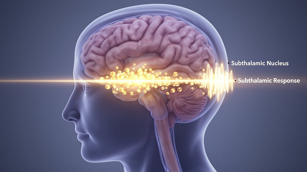
In an intracranial study of 15 people with Parkinson’s disease undergoing deep brain stimulation, dopamine amplified a delayed response in the subthalamic nucleus after motor-cortex stimulation but did not significantly alter the earliest cortical-subthalamic response. The investigators recorded subthalamic local field potentials and cortical electrocorticography while stimulating each region electrically in medication-on and medication-off states. The result identifies a temporal separation within human cortico-subthalamic communication: a fast response consistent with monosynaptic hyperdirect-pathway activation remained comparatively stable, whereas a later response thought more likely to reflect polysynaptic network activity changed with dopamine availability. Because the study involved a small, postoperative cohort, it establishes a circuit-level association in this group rather than a general outcome of treatment for Parkinson’s disease.
The researchers used a bidirectional stimulation-and-recording design during the days after electrodes were implanted. Temporary subdural electrocorticography strips were placed over sensorimotor cortex, while directional deep brain stimulation leads targeted the subthalamic nucleus. Motor cortex was stimulated through a bipolar electrode configuration and responses were recorded from individual subthalamic contacts. In the opposite direction, the researchers stimulated the subthalamic lead and recorded cortical activity. Recordings were repeated with patients taking their usual dopaminergic medication and after withdrawal of dopaminergic medication for at least 12 hours. Clinically meaningful motor improvement accompanied the medication-on state: mean Movement Disorder Society–Unified Parkinson’s Disease Rating Scale Part III score was 25.9 ± 9.7 off medication and 16.6 ± 8.9 on medication.
Cortical stimulation elicited a reproducible series of subthalamic responses. The earliest appeared at 2.6 ± 0.7 milliseconds, followed by components at 10.1 ± 1.9, 23.9 ± 3.4 and 38.9 ± 3.5 milliseconds. The first deflection was significantly different from baseline activity across all patients, with p = 0.002, and its consistent shape and presence across nearly all recorded subthalamic channels supported its interpretation as fast orthodromic engagement of the cortex-to-subthalamus pathway. Its latency and reproducibility were consistent with the hyperdirect projection, which animal and tractography research has associated with a direct, monosynaptic route from cortex to the subthalamic nucleus. The human recordings were compatible with that mechanism, although stimulation artefact or volume conduction could not be excluded completely.
Several checks strengthened the physiological interpretation of the fast response. It occurred after the stimulation artefact ended, retained its form when bipolar stimulation polarity was reversed in one participant, and showed spatial specificity rather than appearing equally across the brain. The researchers excluded the first millisecond, during which the artefact was confined, and defined the earliest analyzable response after the signal returned to baseline. Subthalamic stimulation also produced an early cortical response at 3.2 ± 0.3 milliseconds, followed by later components at 6.4 ± 1.1, 11.5 ± 1.8 and 23.2 ± 2.4 milliseconds. That cortical response differed from baseline at p = 0.003 and was compatible with antidromic activation of cortico-subthalamic axons.
Medication state did not significantly modify these earliest responses. Among 10 paired datasets for cortical stimulation, the short-latency subthalamic response averaged −3.3 ± 1.7 microvolts with dopaminergic medication withdrawn and −2.6 ± 1.4 microvolts on medication, with p = 0.06. Its latency was 2.7 ± 0.6 milliseconds off medication and 2.6 ± 0.8 milliseconds on medication, with p = 0.36. In nine paired datasets for subthalamic stimulation, the short-latency cortical amplitude was −18.2 ± 9.7 microvolts off medication and −22.1 ± 9.8 microvolts on medication, with p = 0.367; latency was 3.1 ± 0.3 and 3.3 ± 0.6 milliseconds, respectively, with p = 0.288. These comparisons did not show significant dopaminergic modulation of the earliest responses in either direction.
The delayed subthalamic response was where the medication effect became clear. Following cortical stimulation, activity between 40 and 100 milliseconds differed significantly from baseline at p = 0.002, and its mean peak latency was 79.9 ± 8.9 milliseconds. With medication withdrawn, the peak was positive at 1.5 ± 1.2 microvolts; with medication present, it was negative at −2.2 ± 2.2 microvolts. The paired comparison in 10 patients yielded p = 0.002, while an unpaired comparison of 13 medication-off and 12 medication-on recordings yielded p < 0.001. A cluster-based permutation test with 1,000 iterations confirmed a more sustained negative response in the medication-on state, principally between 65 and 85 milliseconds. Peak latency itself did not differ significantly between conditions: 81.5 ± 10.2 milliseconds off medication and 73.6 ± 10.7 milliseconds on medication, with p = 0.09.
That effect was direction-specific. The researchers found no significant medication-related change in either early or late cortical responses evoked by subthalamic stimulation. For the late cortical component, amplitude was −3.9 ± 26.8 microvolts off medication and −0.2 ± 24.2 microvolts on medication, with p = 0.059 in the paired analysis of nine datasets; latency was 78.7 ± 10 and 75.5 ± 8 milliseconds, with p = 0.258. A temporally resolved cluster-based permutation test also found no significant effect, with p = 0.736 in the unpaired comparison. The observed asymmetry means that dopamine-sensitive modulation was detected in delayed cortical-to-subthalamic signalling but not in the tested cortical responses to subthalamic stimulation; it does not establish the precise synaptic or cellular mechanism responsible.
Recordings from different contacts added a spatial dimension. Strongest negative responses were concentrated in the dorsolateral subthalamic nucleus, and response amplitude became progressively less negative as distance from a previously defined dorsolateral motor “sweet spot” increased. For the early response, that relationship was supported by a Pearson correlation of r = 0.527 and p = 3.981 × 10⁻⁶. The contact producing the largest short-latency response was also on the same contact level selected for chronic DBS in 12 of 15 patients, or 80% of cases. This exceeded the study’s chance model, which assigned equal probability to four contact levels, with two-sided binomial p < 0.001. The comparison was simplified, individual segment agreement was variable, and the researchers said the finding would require prospective validation before it could guide contact selection.
Motor activity offered another check on the central recordings. Five patients showed peripheral muscle activation after cortical stimulation, detected by surface electromyography and visible contraction. Responses mainly involved contralateral distal upper-limb muscles, especially wrist extension, and began at 24.1 ± 7.1 milliseconds on medication and 28.9 ± 16.4 milliseconds off medication in four paired datasets, with p = 0.75. Their presence did not correspond to systematic changes in the amplitude, morphology or medication modulation of subthalamic potentials, and subthalamic amplitude and latency did not correlate with electromyographic amplitude or latency. This dissociation supports interpreting the subthalamic signals as central cortico-subthalamic responses, but the small motor-response subgroup limits conclusions about anatomical specificity. Overall, the findings distinguish a comparatively dopamine-insensitive fast pathway from dopamine-sensitive delayed network activity in the patients studied, while leaving the routes responsible for the delayed component unresolved.
Subject of Research: Dopaminergic modulation of fast and delayed cortico-subthalamic communication in people with Parkinson’s disease
Article Title: Dopamine amplifies subthalamic long-latency responses to cortical stimulation in patients with Parkinson’s disease
Article References: Beyer, J.-C., Cavallo, A., Vanhoecke, J., Köhler, R. M., Al-Fatly, B., Schneider, G.-H., Faust, K., Brandt, G. A., Kühn, A. A., & Neumann, W.-J. (2026). Dopamine amplifies subthalamic long-latency responses to cortical stimulation in patients with Parkinson’s disease. npj Parkinson's Disease, 12(1), Article 254. https://doi.org/10.1038/s41531-026-01567-6
Image Credits: AI Generated
DOI: 10.1038/s41531-026-01567-6
Keywords: Parkinson’s disease, dopamine, subthalamic nucleus, motor cortex, deep brain stimulation, electrocorticography, basal ganglia, hyperdirect pathway, levodopa, intracranial recordings, amplifies, subthalamic
News Source: Cassandra Pierce. (October 20, 2026). Dopamine amplifies delayed subthalamic responses in Parkinson’s patients. Scienmag.