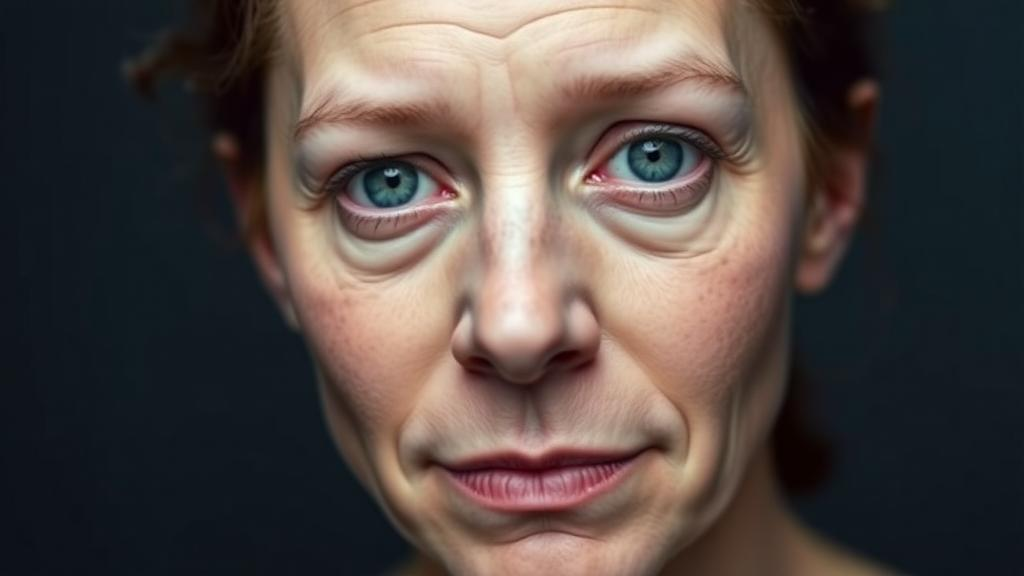
Mass General Brigham researchers have developed a foundation artificial-intelligence model that can estimate biological age and directly predict survival outcomes in patients with cancer from facial photographs. The model-development and validation study, published in The Lancet Digital Health on October 19, 2026, introduces two tools derived from a system trained on more than 40 million face images: FAHR-FaceAge, which estimates biological age, and FAHR-FaceSurvival, which predicts cancer-survival outcomes. The investigators reported that the difference between estimated biological age and chronological age was independently associated with survival, and that combining the two tools improved survival-model fit in an external cohort of cancer patients.
FAHR-Face stands for Foundation Artificial Intelligence for Health Recognition. Its design follows the foundation-model approach: a large system first learns a general representation from a broad collection of face images and is then adapted to particular health-related outcomes. Rather than training an independent algorithm from scratch for each task, the researchers used the shared model to develop both an age estimator and a survival predictor. This structure also allowed them to compare the two outputs directly and ask whether biological-age information and survival information were redundant. The study was computational rather than a clinical trial: it developed and validated prediction models but did not test whether physicians using the scores made better treatment decisions or whether the tools changed patient outcomes.
FAHR-FaceAge was reported to estimate biological age more accurately than previous age-estimation algorithms. The researchers evaluated the model with photographs subjected to common variations encountered in real-world imaging, including differences in pose, lighting, facial expression and makeup. Results remained consistent across most of these conditions, indicating that the age estimates were not restricted to a single photographic setup. The model was also stable across most plastic-surgery procedures included in the analysis. A small shift was observed following rhinoplasty, while the other tested procedures did not produce comparably large changes in the reported estimates. These findings concern the model’s stability under the specific variations and procedures tested; they do not establish that its performance is unaffected by every possible image or medical condition.
FAHR-FaceSurvival takes a different approach from estimating an age-related characteristic and then testing its relationship with survival. It was developed to learn cancer-survival outcomes directly from facial photographs. The age and survival predictions were only weakly correlated, suggesting that the two tools did not simply reproduce the same output. When the measures were analyzed together, each remained independently associated with patient survival. Combining them also improved the fit of the survival model in the external validation cohort of cancer patients. This added model fit is evidence that the two facial-derived measures may contain complementary prognostic information, but the study did not establish a clinical decision threshold, treatment-selection rule or safe way for either score to be used on its own.
Visual analysis of the models showed that they assigned the greatest influence to different parts of a face. FAHR-FaceAge focused on the nasolabial folds, the forehead immediately above the eye sockets and the temples. FAHR-FaceSurvival instead emphasized the area below the eyes and the region around the nasal bridge. The researchers used these maps to show which facial regions most influenced the respective estimates and predictions. The differing regions accompanied the weakly correlated outputs, but the study did not identify a biological mechanism linking particular facial areas to either biological aging or cancer survival. Accordingly, the maps describe the models’ use of visual information rather than proving that the highlighted areas are direct measures of a specific physiological process.
The analysis also identified associations between lifestyle exposures and the biological-age estimates. Heavy alcohol use, drug use and smoking were each associated with looking biologically older. Smoking showed the clearest pattern: current smokers appeared older than former smokers, and former smokers appeared older than people who had never smoked. These were associations observed in the study data rather than evidence that smoking or other behaviors caused the estimated differences or that changing those behaviors would reverse the model’s estimate. The results also do not show that the age tool was intended to diagnose lifestyle exposures. Instead, they indicate that facial-age estimates varied in relation to reported behaviors, alongside variation associated with chronological age.
In examining prognosis, the investigators asked whether the direct survival score and the difference between FAHR-FaceAge and chronological age contributed information beyond one another. Both measures were independently associated with survival when analyzed in the same model, and their combination improved model fit in the external cancer cohort. This supports the interpretation that the age and survival tools can represent related but separable information within the study. It does not mean that a facial photograph alone reveals the cause of a patient’s prognosis, nor does it establish that biological age functions as a mechanism leading to shorter survival. The reported result concerns statistical association, predictive performance and incremental model fit in the populations included in the development and validation work.
Raymond Mak, a radiation oncologist at Mass General Brigham Cancer Institute and faculty member in the institution’s Artificial Intelligence in Medicine program, said the broader goal is to make health assessment from face photographs more quantitative, consistent and scalable. He described this objective in the context of estimating physical reserve and frailty before physicians consider intensive treatments such as radiation, surgery or systemic therapy. The current study, however, did not compare AI-guided treatment selection with standard care or measure safety, treatment response or downstream clinical benefit. Mak and co-senior author Hugo Aerts presented the tools as potential support for personalized treatment decisions rather than replacements for physician judgment. Their findings therefore support further investigation of the scores rather than a demonstrated clinical practice change.
Aerts said FAHR-FaceSurvival advances facial health-recognition research by learning survival risk directly from photographs of patients with cancer. The team described the result as the first demonstration, to its knowledge, of an AI model directly predicting cancer survival from a simple photograph. That direct-learning approach distinguishes the survival tool from an age-estimation model used as an indirect feature. The evidence presented nevertheless remains within the boundaries of a model-development and validation study. No specific biological pathway, patient subgroup in which the tool was safe to deploy, or standard against which AI-assisted care was compared was established in the release.
The researchers are extending the work through the FaceAge Research Study, an online study approved by an institutional review board and open to the public. Participants can upload a facial photograph, receive a FaceAge research readout and contribute data intended to help develop and validate AI-based measures of facial aging and health in larger and more diverse populations. Additional evaluation will be needed to determine how the models perform across broader demographic and clinical settings, beyond the robustness already observed for pose, lighting, expression, makeup and most tested plastic-surgery procedures. The study’s supported conclusion is that FAHR-Face can derive biological-age estimates and direct cancer-survival predictions from facial photographs, with the combined measures adding prognostic value to the survival model in external validation. It is not yet evidence that using those predictions improves treatment choices or clinical outcomes.
Subject of Research: Development and validation of facial-photograph AI models for biological-age estimation and cancer-survival prediction
Article Title: AI facial health recognition tools help physicians assess age and survival risk to guide personalized treatment
Article References: AI facial health recognition tools help physicians assess age and survival risk to guide personalized treatment. Original publication (Bibliographic details unavailable.)
Image Credits: AI Generated
DOI: Not provided
Keywords: facial artificial intelligence, biological age, cancer survival, foundation models, computer vision, medical imaging, prognostic modeling, external validation, digital health, patient photographs, facial, health
News Source: Nathaniel Bowman. (October 20, 2026). Facial AI estimates biological age and predicts cancer survival. Scienmag.