When arteries supplying the legs narrow progressively, the muscle downstream is starved of oxygen and nutrients, and the damage reaches all the way down to the cellular power plants. A new study published in the Journal of Cellular and Molecular Medicine suggests that this damage has a hard limit: once peripheral arterial disease advances to its most severe stage, the mitochondria in the affected muscle appear to lose their capacity to adapt and recover, even after surgeons successfully restore blood flow. The finding, reported by researchers at Charité-Universitätsmedizin Berlin, could reshape how clinicians think about the timing of intervention in a disease that affects hundreds of millions of people worldwide.
Peripheral arterial disease, or PAD, is typically stratified by severity. In its milder form, known as intermittent claudication, patients experience cramping pain during walking that resolves with rest. At the opposite end of the spectrum lies chronic limb-threatening ischemia, or CLTI, defined by persistent rest pain, non-healing wounds, and critically low perfusion measures such as an ankle brachial index below 0.4. Progression from claudication to CLTI is driven by diabetes, hypertension, smoking and ageing, and it is accompanied by profound molecular changes in skeletal muscle. Repeated cycles of ischemia and reperfusion impair cellular metabolism, intensifying both mitochondriopathy and myopathy as the disease advances, while progressive fibrosis and altered gene expression set severely affected muscles apart from healthier tissue.
The research team hypothesised that the adaptive capacity of mitochondrial respiration after successful revascularisation would be impaired in advanced disease stages. To test this, they enrolled 32 consecutive patients with symptomatic PAD who required treatment: 21 with intermittent claudication and 11 with CLTI. The two groups were comparable in age, sex distribution, body mass index, comorbidities and medication. Among the CLTI patients, most suffered from rest pain or chronic wounds, and all eight patients with wounds showed improved healing after surgery, with seven achieving complete resolution. Revascularisation was performed by open surgery, endovascular techniques, or a hybrid of both, and its success was confirmed by duplex ultrasound together with significant improvements in ankle brachial index and walking distance in both groups.
The methodological heart of the study lies in high-resolution respirometry, a technique that measures oxygen consumption in living mitochondria with extraordinary precision. Using a Bergstrom biopsy needle, the researchers took small tissue samples from two muscles of the affected leg: the gastrocnemius, the calf muscle that lies directly downstream of the typical arterial blockages, and the vastus lateralis of the thigh, which serves as a less affected internal reference. Biopsies were taken perioperatively and again six weeks after revascularisation. The muscle fibres were chemically permeabilised with saponin, allowing substrates and inhibitors direct access to the mitochondria, and then transferred to the respirometry chambers of an Oxygraph-2k instrument maintained at 37 degrees Celsius.
Inside the chambers, the researchers followed a substrate-uncoupler-inhibitor titration protocol that maps the entire functional landscape of the respiratory chain. Malate and octanoylcarnitine first induced LEAK respiration, the resting state without ADP. Adding ADP activated Complex I-linked oxidative phosphorylation, and further titration of pyruvate and succinate revealed the combined capacity of Complexes I and II working together. Stepwise addition of the uncoupler FCCP pushed the system to maximal electron transfer capacity, exposing the upper limit of respiratory chain activity, before rotenone shut down Complex I to isolate Complex II respiration. Because mitochondrial content varies between individuals, the team also measured citrate synthase activity, a standard marker of mitochondrial abundance, and normalised respiration values accordingly.
The results revealed a striking stage-dependent pattern. In the claudication group, mitochondrial respiration per unit of citrate synthase activity in the calf muscle declined significantly after revascularisation, particularly for maximal ADP-stimulated respiration and for Complex II activity measured after Complex I inhibition. Compared with control values from the team’s previous work, this decline represented a normalisation: preoperatively elevated respiration dropped back toward healthy levels once blood flow was restored. In the CLTI group, by contrast, no significant changes appeared in any respiratory state six weeks after surgery. The severely ischemic muscles simply did not respond to the renewed oxygen supply, even though the intervention itself was clinically successful.
An elegant internal control reinforced this conclusion. Because high-resolution respirometry in PAD patients shows considerable inter-individual variability, the researchers calculated a gastrocnemius-to-vastus ratio, comparing respiration in the ischemic calf muscle against the same patient’s thigh muscle. In the claudication group, this ratio decreased significantly after revascularisation for ADP-stimulated respiration, oxidative phosphorylation capacity, electron transfer capacity and Complex II respiration, again suggesting a return toward normal physiology. The CLTI group showed no significant changes at all. Interestingly, citrate synthase activity did rise postoperatively in the calf muscle of CLTI patients, possibly reflecting increased physical activity after surgery, but this increase in mitochondrial quantity was not accompanied by any measurable improvement in mitochondrial quality or function.
Why would mitochondria in severely ischemic muscle lose their adaptability? The authors point to several converging mechanisms. Dysfunctional mitochondria produce excess reactive oxygen species, and once the burden of oxidative stress exceeds a critical threshold, the protective phenomenon of mitohormesis, in which mild stress stimulates antioxidant defences, appears to fail. More pronounced ischemia-reperfusion injury in CLTI further damages mitochondrial machinery and reduces the potential for cellular repair. Impaired microcirculation, which may be only partially reversible even after the macroscopic vessels are reopened, likely limits tissue-level adaptation as well. Exercise training, the standard adjunct therapy, primarily enhances mitochondrial content rather than function, and therefore cannot compensate for these deeper deficits. Genomic and proteomic analyses by other groups support the picture of CLTI as a biologically distinct entity, with enriched fibrosis pathways and a downregulated mitochondrial proteome in amputation-stage patients.
The clinical implications are significant. Adequate reperfusion does not always translate into clinical improvement, and this study offers a mechanistic explanation: in advanced disease, the window for mitochondrial recovery may already have closed. The findings underscore the importance of early diagnosis and timely intervention, before the adaptive capacity of the muscle is exhausted. They also raise the question of whether patients with CLTI might need additional, mitochondria-targeted therapies alongside revascularisation, or whether longer follow-up periods might reveal delayed regenerative responses that six weeks are too short to capture. An ongoing randomised controlled trial is investigating whether surgical revascularisation or supervised exercise therapy better promotes mitochondrial adaptation in PAD, work that could shape future personalised treatment paradigms.
The study has limitations that the authors acknowledge candidly. The CLTI cohort was small, particularly for postoperative biopsies, a reflection of the frailty of these patients and their reluctance to undergo repeated invasive sampling. Only one mitochondrial content marker was available due to limited tissue, and the ankle brachial index may overestimate perfusion in diabetic patients with calcified vessels. There was no matched control group without PAD, and no data on physical activity patterns during the six-week recovery period. Nevertheless, the central message stands: the adaptability of mitochondrial function after revascularisation appears largely confined to earlier stages of peripheral arterial disease. For clinicians, the study is a reminder that restoring blood flow is necessary but not always sufficient, and that the biology of the muscle itself determines whether recovery is possible.
Subject of Research: Mitochondrial adaptability in skeletal muscle after revascularisation in peripheral arterial disease
Article Title: Chronic Limb‐Threatening Ischemia Shows No Adaptability of Mitochondrial Function After Revascularisation
Article References: Speichinger, F., Gratl, A., Raude, B., Arndt, M., Hering, N. A., Greiner, A., Pesta, D., Schineis, C., & Frese, J. P. (2026). Chronic Limb‐Threatening Ischemia Shows No Adaptability of Mitochondrial Function After Revascularisation. Journal of Cellular and Molecular Medicine, 30(20), Article e71383. https://doi.org/10.1111/jcmm.71383
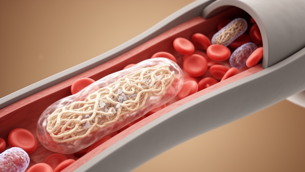
Image Credits: AI Generated
DOI: 10.1111/jcmm.71383
Keywords: peripheral arterial disease, chronic limb-threatening ischemia, mitochondria, revascularisation, high-resolution respirometry, intermittent claudication, skeletal muscle, citrate synthase, ischemia-reperfusion injury, vascular surgery, oxidative phosphorylation, mitohormesis
News Source: Drew Townsend. (October 19, 2026). Mitochondria in Severe Leg Artery Disease Lose Their Ability to Recover After Revascularisation. Scienmag.