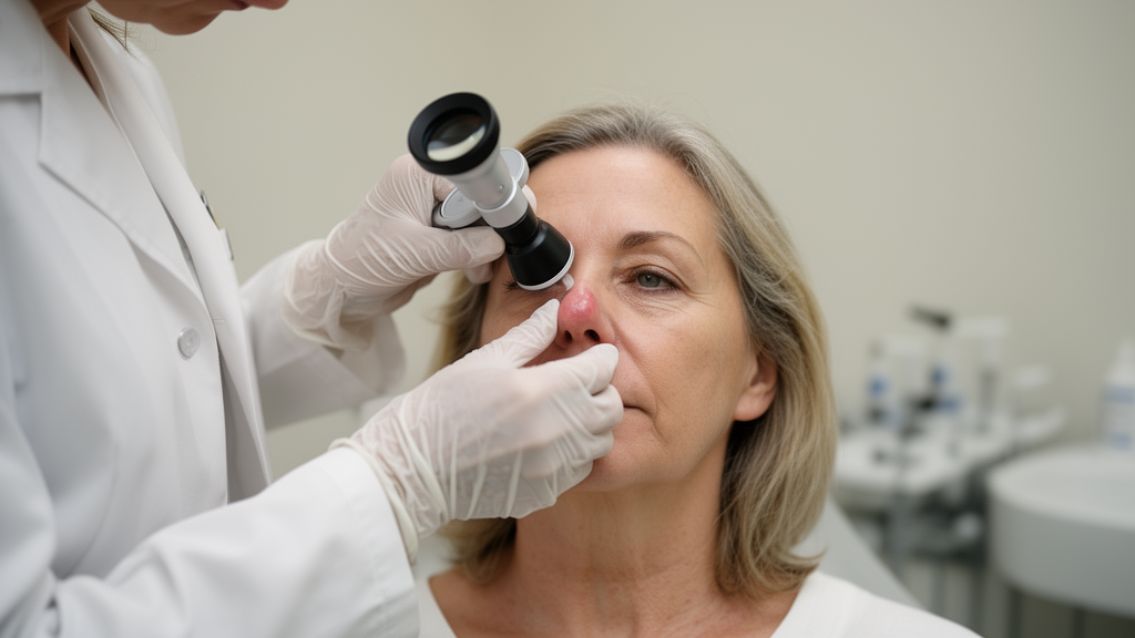
A small lump on the side of a 63-year-old woman’s nose looked, to every clinical eye that examined it, like a lipoma — the utterly ordinary fatty bump that dermatologists and surgeons encounter thousands of times a year. It was painless, well circumscribed, slowly enlarging, and showed none of the dramatic symptoms that usually announce a tumor. But when surgeons at Vilnius University in Lithuania excised the mass and pathologists placed it under the microscope, the diagnosis was anything but ordinary: the lesion was a glomangiomyoma, an exceptionally rare variant of a glomus tumor arising in the superficial soft tissues of the nose. To the authors’ knowledge, it is the first such case ever reported in Lithuania, and one of only a handful of superficial facial glomangiomyomas described anywhere in the world.
Glomus tumors are uncommon benign neoplasms of the pericytic family — tumors built from modified smooth muscle cells of the glomus body, a specialized arteriovenous structure involved in thermoregulation. The glomus body is essentially a biological thermostat valve: a direct connection between arteries and veins wrapped in contractile cells that can open or close blood flow through the skin in response to temperature. These structures are densest in the fingers and toes, which is why roughly 1.6% of all soft tissue tumors are glomus tumors and why the overwhelming majority occur in the subungual regions of the extremities, beneath the fingernails and toenails. A glomus tumor under a nail is hard to miss: it produces severe pain, pinpoint tenderness, and a notorious cold sensitivity that patients describe as excruciating.
Facial glomus tumors are a different beast entirely. According to the focused literature review conducted by the Lithuanian team and published in Frontiers in Medicine, reported superficial facial lesions have involved the cheek, lip, eyelid, forehead, and nasal soft tissues — but the number of documented cases is vanishingly small. More importantly, many of these facial tumors lack the characteristic pain of their digital counterparts. In a 20-year series of 56 extradigital glomus tumors published by Schiefer and colleagues at the Mayo Clinic, cold hypersensitivity occurred in only 2% of patients, and the mean diagnostic delay exceeded seven years. Only one nasal and one cheek lesion were identified in that entire two-decade experience. Smith and colleagues, reviewing 31 oral and paraoral glomus tumors, found the lips to be the most frequent site and reported pain or tenderness in fewer than half of cases.
The present case illustrates precisely why this matters. The woman’s lesion had first been noticed approximately three years earlier as a small subcutaneous mass in the upper right nasal sidewall. It grew gradually but was never painful, never tender, never sensitive to cold, and never associated with bleeding, skin discoloration, or any other local symptom. Clinical examination revealed a small, well-circumscribed subcutaneous mass without overlying skin changes or signs of inflammation. Because it appeared clinically benign and small, no preoperative imaging was performed at all. The working diagnosis was lipoma, and surgical excision under local anesthesia was undertaken for both diagnostic and therapeutic purposes — a routine decision that would uncover something no one expected.
The excised specimen consisted of a grayish soft-tissue fragment measuring just 0.7 by 0.5 by 0.4 centimeters. Histopathological examination revealed a well-circumscribed subcutaneous neoplasm composed of variably sized vascular spaces surrounded by uniform rounded epithelioid glomus cells with small round nuclei and eosinophilic cytoplasm. But the tumor contained a second population of cells that told a deeper story: intermixed with the epithelioid glomus cells were fascicles of spindle-shaped smooth-muscle cells with abundant eosinophilic cytoplasm and elongated, cigar-shaped nuclei. Crucially, the pathologists identified a gradual morphological transition between the two components — rounded glomus cells slowly giving way, cell by cell, to mature smooth-muscle cells. That transition is the morphological signature of glomangiomyoma, a distinct entity recognized by the 2020 World Health Organization classification of soft tissue tumors within the pericytic (perivascular) tumor group.
Immunohistochemistry — the technique of using labeled antibodies to detect specific proteins in tissue sections — confirmed the diagnosis with elegant precision. The glomus cells demonstrated diffuse cytoplasmic positivity for smooth muscle actin (SMA), consistent with their perivascular myoid differentiation. CD34, an endothelial marker, highlighted the lining of the vascular channels rather than the neoplastic cells themselves. The spindle-shaped smooth-muscle component showed strong desmin positivity, while the rounded glomus cells were desmin-negative — a clean immunohistochemical split that mirrored the morphological transition. The Ki-67 proliferation index, a measure of cell division, was below 1%, and there was no significant cytological atypia, tumor necrosis, or increased mitotic activity. Together, these findings painted a picture of a benign tumor with a very low proliferative rate — but one whose classification required careful attention to both morphology and immunohistochemistry.
The distinction matters technically. SMA expression is characteristic of glomus tumors more broadly, and by itself does not establish the diagnosis of glomangiomyoma. What separates glomangiomyoma from conventional glomus tumor is the morphological evidence of smooth-muscle differentiation — the gradual transition from conventional rounded glomus cells to elongated spindle-shaped cells exhibiting mature smooth-muscle features. The histopathological differential diagnosis also includes other perivascular and smooth-muscle neoplasms, particularly myopericytoma and angioleiomyoma. Conventional glomus tumors are characterized by relatively uniform rounded cells arranged around vascular channels, whereas the conspicuous transition seen here supported glomangiomyoma. The authors also caution against a common historical confusion: head and neck paragangliomas were once also called glomus tumors, but they represent an entirely distinct neuroendocrine neoplasm and should not be confused with true soft-tissue glomus tumors.
The surgical outcome was textbook. The lesion was completely excised, and histopathological examination confirmed that the closest resection margin was free of tumor, with a microscopic clearance of 0.5 millimeters. The postoperative course was uneventful, and at six-month clinical follow-up the surgical site was well healed with no evidence of local recurrence or postoperative complications. Complete surgical excision is the treatment of choice for localized superficial glomus tumors, with recurrence reported predominantly in association with incomplete excision. However, the authors are careful to note that six months is a relatively short follow-up period that does not permit conclusions about long-term recurrence, and the available facial literature is limited predominantly to individual case reports and small series with heterogeneous, frequently incompletely reported follow-up.
The literature review conducted by the team — searching PubMed, Scopus, and Google Scholar with terms including glomus tumor, glomangioma, glomangiomyoma, facial, cheek, lip, nasal, forehead, chin, eyelid, and periorbital, last updated in September 2026 — identified a strikingly limited number of reported superficial facial glomus tumors. Among the documented cases were a 50-year-old woman with a facial glomus tumor presenting with pain on accidental touch, a 24-year-old man with a painful cheek lesion that recurred two months after excision, a series of seven children and young adults with asymptomatic facial glomangiomas involving the cheek, lips, and lower eyelid, and a 25-year-old man with an asymptomatic facial glomangiomyoma that showed no recurrence at 23 months. A 28-year-old man described in 2026 had a painless superficial cheek glomus tumor initially misclassified on ultrasonography and diagnosed definitively only after histopathological and immunohistochemical evaluation. One patient in the review had carried an asymptomatic lesion for 30 years before diagnosis.
The principal clinical value of this case, the authors argue, is not rarity alone but the potential for an extradigital facial glomus tumor to mimic a common benign soft-tissue lesion. The painless, clinically innocuous appearance of the nasal mass led to an initial suspicion of lipoma, with the definitive diagnosis established only after excision and histopathological examination. The message for clinicians is clear: glomus tumors should be considered in the differential diagnosis of persistent superficial facial nodules even when the characteristic pain, pinpoint tenderness, and cold sensitivity of digital lesions are entirely absent. Histopathological examination remains essential for definitive diagnosis, and in uncommon subtypes such as glomangiomyoma, careful assessment of cellular morphology supported by appropriate immunohistochemistry is necessary for accurate classification. For a lump that looked like the most boring bump in medicine, the story it told under the microscope was one of the rarest in facial pathology.
Subject of Research: Medicine
Article Title: Nasal glomangiomyoma: a case report and review of superficial facial glomus tumors
Article References: Kindurytė-Šepetienė, A., Glovackaitė, P., Markevičius, D., Šidlovska, E., Stundžaitė-Baršauskienė, G., & Jakutis, N. (2026). Nasal glomangiomyoma: a case report and review of superficial facial glomus tumors. Frontiers in Medicine, 13, Article 1960678. https://doi.org/10.3389/fmed.2026.1960678
Image Credits: AI Generated
DOI: 10.3389/fmed.2026.1960678
Keywords: glomus tumor, glomangiomyoma, nasal tumor, facial soft tissue, pathology, case report, pericytic neoplasm, immunohistochemistry, Nasal, case, report, review
News Source: Ophelia Keating. (October 19, 2026). Rare Painless Tumor on a Woman’s Nose Turns Out to Be Something Far Stranger. Scienmag.