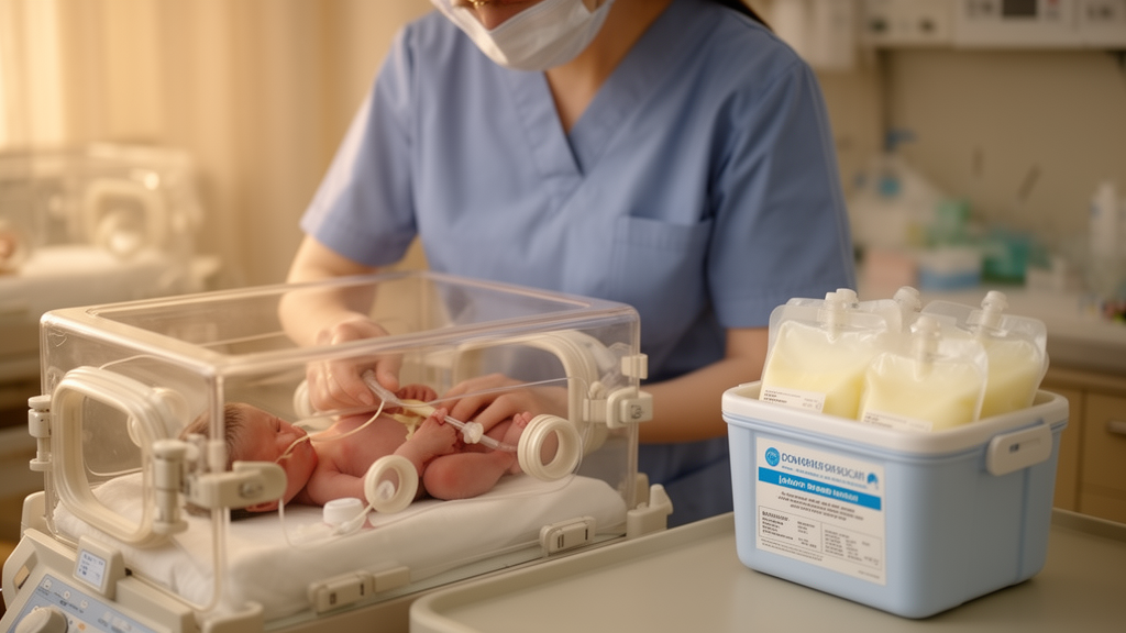
For the smallest and most fragile newborns, every milliliter of milk and every day of nutrition carries enormous weight. Babies born before 32 weeks of gestation, often weighing little more than a kilogram, cannot yet coordinate sucking and swallowing, so their first feeds are delivered through a thin tube directly into the stomach. The question of what should go into that tube—mother’s own milk, pasteurized donor human milk, or a blend of the two—has long divided neonatal intensive care units, even as donor milk programs have expanded dramatically across the United States. A new study now offers a strikingly reassuring answer: when it comes to the earliest and most critical feeding milestones, the source of the human milk may not matter as much as clinicians and parents have feared.
The research, published in the Journal of Perinatology, was led by nutrition scientist Lyssa Lamport of Rutgers University together with colleagues at Cohen Children’s Medical Center of New York. The team carried out a retrospective cross-sectional chart review of 359 very low birth weight infants born at less than 32 weeks gestation between January 2021 and December 2024. Their goal was to determine whether the type of human milk an infant received during the earliest phase of enteral nutrition influenced three key measures of neonatal feeding progress: the number of days until human milk fortification was added, the number of days until full enteral nutrition was achieved, and the total duration of parenteral nutrition, in which nutrients are delivered intravenously.
The stakes of these milestones are far from abstract. Human milk is widely regarded as the gold standard for feeding premature infants, and an exclusive human milk diet has been linked to significant reductions in prematurity-related complications such as necrotizing enterocolitis, a potentially devastating bowel disease, and late-onset sepsis, a bloodstream infection. Reaching full enteral nutrition as quickly and safely as possible is also essential because prolonged reliance on intravenous nutrition carries its own risks, including infection and liver injury. Feeding milestones and parenteral nutrition duration therefore serve as critical indicators of an infant’s maturation and overall development.
Yet mother’s own milk and donor milk are not biologically identical. Mother’s own milk, particularly in the earliest days after birth, is a dynamic biological fluid packed with bioactive components that support immune protection and intestinal maturation. Colostrum, the first milk produced after delivery, contains the highest concentration of these factors, which gradually decline as milk matures. Donor milk, by contrast, is typically collected from mothers of older infants and contains lower levels of protein, fat, and bioactive components. The Holder pasteurization process required for safety further reduces immune cells, anti-inflammatory factors, and beneficial bacteria, leaving mainly heat-stable components at diminished concentrations. These disparities have raised persistent questions about whether donor milk can truly substitute for a mother’s own colostrum and milk during the most vulnerable window of a preterm infant’s life.
The urgency of answering that question has grown alongside the explosive expansion of donor milk use. In advanced-level neonatal care units in the United States, donor milk utilization rose from 28.7 percent in 2009 to 54.7 percent in 2020, and by 2024 it exceeded 95 percent. Many units now initiate early enteral feeding with donor milk while waiting for a mother’s milk supply to come in, a practice designed to avoid formula but one that may inadvertently limit a newborn’s exposure to colostrum. Despite this widespread adoption, few studies have directly compared exclusive mother’s own milk, exclusive donor milk, and mixed feeding regimens, largely because infants in donor milk groups are usually also fed some maternal milk, making it impossible to isolate the effects of an exclusively donor milk diet.
The New York team took advantage of a setting in which the two milks were fed separately and meticulously recorded, allowing exact calculation of each infant’s intake. They classified infants into four groups based on the proportion of mother’s own milk versus donor milk received from the first enteral feed until fortification: 100 percent mother’s own milk, 100 percent donor milk, donor milk with at least 50 percent maternal milk, and donor milk with less than 50 percent maternal milk. The study period for this classification deliberately ended at the time of fortification, providing a standardized window for evaluating early, unfortified milk exposure. Infants who had received any formula, who had not begun enteral feeding within 72 hours of life, or who had developed serious bowel complications before the first feed were excluded, along with infants transferred in after 24 hours of life and those with congenital anomalies or metabolic disorders.
The cohort was typical of modern neonatal intensive care populations. Median gestational age was 28.9 weeks, mean birth weight was about 1060 grams, and slightly more than half the infants were female. The first enteral feed was initiated on average at roughly 36 hours of life, and nearly half of the infants received mother’s own milk as that first feed. About 30 percent received exclusive maternal milk during the study window, 11 percent received exclusive donor milk, and the remaining 59 percent received a mixture. Across the whole cohort, fortification was added at a median of five days, full enteral nutrition was reached at nine days, and parenteral nutrition lasted a median of ten days.
When the researchers analyzed the data using a generalized linear model with a negative binomial distribution, appropriate for overdispersed count data, and adjusted for birth weight, five-minute APGAR score, intrauterine growth restriction, and the hour of the first feed, the results were unambiguous. No statistically significant differences emerged among the milk type groups for days to fortification, days to full enteral nutrition, or days of parenteral nutrition. The P values ranged from 0.70 to 0.79, far from the threshold of significance. This finding directly contradicted the team’s original hypothesis that starting with 100 percent donor milk would delay feeding milestones and prolong intravenous nutrition.
The unadjusted data told a more complicated and instructive story. Before statistical correction, infants fed exclusively mother’s own milk appeared to take longer to reach fortification, full feeding, and independence from parenteral nutrition, and they had the longest hospital stays. But that group also had the lowest mean birth weight, at about 989 grams, and a higher rate of low five-minute APGAR scores, suggesting they were simply smaller and sicker from the start. Previous research has shown that each one-week decrease in gestational age, each 100-gram drop in birth weight, or each point lower on the five-minute APGAR score raises the likelihood of delayed feeding advancement by eight to twenty percent. Once these confounders were accounted for, the apparent disadvantage of maternal milk vanished, underscoring how profoundly an infant’s underlying condition shapes feeding outcomes regardless of milk source.
The authors emphasize that their findings offer practical reassurance for clinicians and families alike: prioritizing human milk feeding, including an exclusive donor milk diet, does not impede feeding progression or extend the need for intravenous nutrition. The results reinforce recommendations from the American Academy of Pediatrics and the World Health Organization to maximize human milk feeding in preterm infants, using donor milk when maternal milk is insufficient or unavailable, and they strengthen the case for integrating donor milk programs into standard neonatal protocols. At the same time, the study leaves important questions open. A compelling but statistically underpowered trend showed that infants receiving higher proportions of maternal milk were more likely to receive colostrum before tube feeding began and had the lowest rates of necrotizing enterocolitis, consistent with a dose-dependent protective effect. Because the study was retrospective and cross-sectional, causality cannot be established, and it examined only short-term feeding milestones rather than growth, neurodevelopment, or long-term immune outcomes. The authors call for larger, adequately powered prospective studies to determine whether an exclusive donor milk diet, in the absence of early colostrum exposure, might still compromise protection against necrotizing enterocolitis and to explore how milk type interacts with degrees of prematurity and growth restriction. For now, the central message stands: in the race to full feeding, what matters most may be that preterm infants receive human milk at all.
Subject of Research: Effects of mother's own milk versus donor human milk on neonatal feeding milestones in preterm infants
Article Title: Early enteral nutrition with mothers’ own milk, donor human milk, or a combination of both on neonatal feeding milestones: a cross-sectional study
Article References: Lamport, L., Samavat, H., Gomes, A., Newkirk, M., Byham-Gray, L., & Ziegler, J. (2026). Early enteral nutrition with mothers’ own milk, donor human milk, or a combination of both on neonatal feeding milestones: a cross-sectional study. Journal of Perinatology. https://doi.org/10.1038/s41372-026-02928-y
Image Credits: AI Generated
DOI: 10.1038/s41372-026-02928-y
Keywords: preterm infants, donor human milk, mother's own milk, enteral nutrition, parenteral nutrition, human milk fortification, necrotizing enterocolitis, very low birth weight, neonatal intensive care, colostrum, feeding milestones, Journal of Perinatology
News Source: Harold Sullivan. (October 19, 2026). Donor Milk Matches Mother’s Own Milk for Preterm Feeding Milestones, Study Finds. Scienmag.