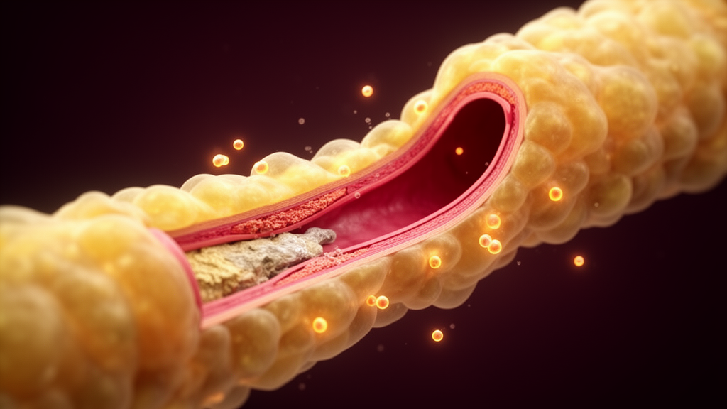
Scientists have found a way to read inflammation directly from the fat that surrounds the heart’s most critical artery, and the numbers they extracted turned out to be strikingly informative. In a new study published in Frontiers in Cardiovascular Medicine, a research team in Kashi, China, reports that a CT-derived measurement called the perivascular fat attenuation index, or FAI, is strongly associated with anatomically significant narrowing of the left anterior descending coronary artery, the vessel famously nicknamed the widow-maker for its role in fatal heart attacks. When the team combined this fat-based signal with three blood biomarkers, the resulting diagnostic model distinguished patients with severe artery narrowing from those with milder disease with an apparent accuracy approaching 92 percent, a performance level that has generated considerable excitement in the cardiovascular imaging community.
The biology behind the measurement is as elegant as it is counterintuitive. Fat tissue wrapped around coronary arteries is not a passive bystander; it behaves like a biological thermostat for the vessel wall beneath it. When a coronary artery becomes inflamed by atherosclerosis, inflammatory signaling molecules released from the diseased vessel wall diffuse outward into the adjacent perivascular fat, where they suppress the formation of new fat cells, promote lipolysis, and increase the water content of the tissue. Because CT scanners measure tissue density in Hounsfield units, and because fat is normally very dense in the negative direction, this inflammatory waterlogging makes the fat appear less negative on the scan. A higher, less negative FAI value therefore serves as a noninvasive fingerprint of local coronary inflammation, something no conventional angiogram can reveal.
To test whether this fingerprint tracks real artery disease, the researchers retrospectively analyzed 160 patients with suspected coronary artery disease who had undergone coronary computed tomography angiography, or CCTA, at The First People’s Hospital of Kashi Prefecture between January 2019 and February 2021. The cohort was deliberately purified: patients on statins or any anti-inflammatory medications, including corticosteroids, chronic nonsteroidal anti-inflammatory drugs, or colchicine, were excluded to prevent treatment from distorting the inflammatory signals being measured. Patients with prior heart attacks, revascularization, cardiomyopathy, significant disease in coronary vessels other than the LAD, acute infection, systemic inflammatory disease, or malignancy were also excluded. After screening, 78 patients had LAD stenosis below 50 percent and 82 had anatomically significant stenosis of at least 50 percent, the prespecified primary outcome.
Quantifying FAI required careful, standardized image processing. Using an artificial intelligence-assisted post-processing platform, the team generated a three-dimensional volume of interest around a 40-millimeter segment of the LAD centered on the point of maximal narrowing, extending 20 millimeters in each direction. The software identified perivascular adipose tissue within a CT window of minus 190 to minus 30 Hounsfield units, and the sampling radius extended from the external vessel wall outward by a distance equal to the local vessel diameter, consistent with the internationally recognized CRISP-CT methodology. Structures such as the aortic root, left atrial appendage, pulmonary trunk, and any calcified plaque were manually excluded. Two experienced cardiovascular radiologists, blinded to all clinical and biomarker data, reviewed the segmentations, and reproducibility testing in a dual-read subgroup of 40 scans showed an intraclass correlation coefficient of 0.991, indicating that the measurement is highly consistent between readers.
The results were unambiguous. Patients with anatomically significant LAD stenosis had a mean FAI of minus 64.58 Hounsfield units, considerably less negative than the minus 80.10 units observed in patients with milder narrowing, a difference that was highly statistically significant. FAI correlated with stenosis severity both as a continuous percentage, with a Spearman rank correlation coefficient of 0.54, and as a binary threshold at 50 percent, where the correlation rose to 0.64. Notably, the mean FAI in the severe stenosis group sat above the minus 70.1 Hounsfield unit threshold that the landmark CRISP-CT study previously linked to elevated cardiac mortality, while the milder group sat below it, although the authors caution that this comparison is descriptive only because the CRISP-CT threshold was derived in a different cohort with a different endpoint.
The blood biomarkers told a complementary story. Patients with severe narrowing had markedly higher levels of high-sensitivity C-reactive protein, the classic circulating marker of low-grade systemic inflammation, with median values of 15.50 milligrams per liter compared with 6.87 in the milder group. Lactate dehydrogenase, an enzyme linked in some cohorts to adverse cardiovascular outcomes, was also elevated at 241.30 units per liter versus 183.03. Meanwhile, the apolipoprotein A1 to apolipoprotein B ratio, which captures the balance between antiatherogenic and atherogenic lipoprotein activity, was substantially lower in the severe group at 0.77 versus 1.26. In multivariable logistic regression, all four markers emerged as independent predictors: FAI, hs-CRP, and LDH each carried a positive association with severe stenosis, while the ApoA1/ApoB ratio was inversely associated, and conventional measures such as total cholesterol and HDL did not remain significant after adjustment.
The composite diagnostic model built from these four predictors achieved an area under the receiver operating characteristic curve of 0.915, with a 95 percent confidence interval of 0.865 to 0.965, corresponding to a sensitivity of 88.5 percent and a specificity of 91.2 percent at the optimal cutoff. Formal DeLong testing for correlated ROC curves confirmed that the composite model discriminated significantly better than hs-CRP alone, with a P value of 0.020, and dramatically better than FAI alone, with a P value below 0.001. Internal validation was unusually rigorous for a study of this size: the model was evaluated with stratified 5-fold cross-validation, 10-fold cross-validation, and 1,000-bootstrap resampling, yielding validation AUCs of 0.872, 0.881, and 0.895 respectively, and the entire model-building procedure was repeated inside each resample to avoid the optimism that arises when a model is selected and validated on the same data.
The authors are refreshingly candid about what the model cannot yet do. Because the study was single-centre, retrospective, and cross-sectional, with no follow-up outcome data, no invasive confirmation of ischemia, and no external validation cohort, the findings should be regarded as preliminary and hypothesis-generating rather than clinically actionable. The cohort is also highly selected: only 160 of 513 screened patients were analyzed, patients with multivessel disease, who carry the highest event risk, were systematically absent, and the prevalence of severe stenosis in the analyzed cohort, at 51.2 percent, far exceeds what would be seen in an unselected CCTA population, meaning the reported accuracy figures cannot be extrapolated to routine clinical practice. Bootstrap analysis also revealed that the LDH component was retained in only about half of replicates, marking it as the least stable element of the model, and the optimal FAI threshold was poorly determined, with a 95 percent interval spanning more than 40 Hounsfield units.
What makes the study compelling nonetheless is the way it fuses two complementary windows onto the same disease process. Conventional CCTA defines the anatomy, revealing where and how severely a coronary artery is narrowed, while FAI reads the active vascular biology of the fat wrapped around that artery, and serum biomarkers capture the systemic inflammatory and metabolic milieu in which the plaque is developing. The finding that hs-CRP carried most of the discriminative information in this high-prevalence cohort, with the imaging-derived FAI adding a statistically significant but modest increment, raises an important question for the field: in a population with a more balanced case mix, where clinical judgment is genuinely uncertain, the added value of imaging the perivascular fat space may prove larger than these figures suggest. Testing that hypothesis will require prospective, multicentre cohorts with outcome data, and ideally serial measurements that can establish whether the FAI signal changes as plaques evolve.
For now, the study stands as a vivid demonstration that the fat around our arteries is talking, and that modern CT scanners, paired with AI-assisted analysis and a handful of blood tests, can begin to listen. If the composite approach survives external validation, it could eventually help clinicians identify which patients with chest pain harbor not just narrowed arteries but biologically active, inflamed plaques, the kind most likely to rupture and cause heart attacks. Until then, the widow-maker’s surrounding fat has offered science a new and provocative line of evidence, one that transforms a routine CT scan into a potential window on the inflammatory storm driving coronary disease.
Subject of Research: Association between the perivascular fat attenuation index and CCTA-defined anatomically significant left anterior descending coronary artery stenosis: relationship with serum inflammatory biomarkers
Article Title: Association between the perivascular fat attenuation index and CCTA-defined anatomically significant left anterior descending coronary artery stenosis: relationship with serum inflammatory biomarkers
Article References: Ahongjiang, Y., Abudukaiyoumu, A., Abulimiti, G., Tuerxun, H., & Mutailifu, A. (2026). Association between the perivascular fat attenuation index and CCTA-defined anatomically significant left anterior descending coronary artery stenosis: relationship with serum inflammatory biomarkers. Frontiers in Cardiovascular Medicine, 13, Article 1823667. https://doi.org/10.3389/fcvm.2026.1823667
Image Credits: AI Generated
DOI: 10.3389/fcvm.2026.1823667
Keywords: Association, between, perivascular, attenuation, index, CCTA-defined, anatomically, significant, left, anterior, descending, coronary
News Source: Ophelia Keating. (October 19, 2026). Association between the perivascular fat attenuation index and CCTA-defined anatomically significant left anterior descending coronary artery stenosis: relationship with serum inflammatory biomarkers. Scienmag.