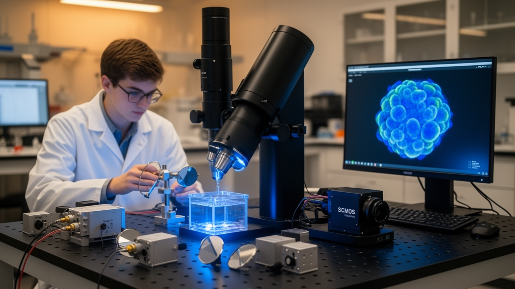
Light-sheet fluorescence microscopy has long promised a gentler, faster way to watch biology unfold in three dimensions, but the field has quietly suffered from a fragmentation problem. Instruments built to capture the flicker of clathrin-coated pits inside a single cell are fundamentally different from those designed to sweep through an entire cleared mouse brain. Now, a team led by researchers at UT Southwestern Medical Center and Calico Life Sciences has introduced a platform that aims to collapse that divide into a single, reconfigurable machine. Writing in Nature Methods, Bingying Chen, Alfred Millett-Sikking, Reto Fiolka and colleagues present the multi-immersion oblique plane microscope, or miOPM, a system that lets scientists swap objectives freely and adapt to samples ranging from living cells to whole organisms and chemically cleared tissue.
The core problem the team set out to solve is architectural. In a conventional light-sheet microscope, illumination and detection objectives sit at right angles to one another, and each pair must be carefully matched and mechanically coordinated. High-resolution variants such as lattice light-sheet microscopy use custom illumination objectives squeezed into the remaining space around the detection lens, a constraint that caps how much emitted light can be collected and makes swapping objectives essentially impossible. The consequence is that a lab studying subcellular dynamics in single cells and one imaging whole embryos typically need entirely different instruments, each demanding its own expertise, mounting hardware and maintenance budget.
Oblique plane microscopy, the approach behind miOPM, sidesteps the orthogonal geometry by using a single objective for both illumination and detection. A light sheet is launched at an angle through the same lens that collects the fluorescence, and a set of downstream optics maps the tilted image plane onto a camera without distortion. The catch, until now, has been that those downstream optics had to be precisely matched to the primary objective. Change the objective and you effectively had to rebuild the microscope. That rigidity has kept OPM powerful but narrow, excellent for adherent cells in dishes but poorly suited to large or non-aqueous specimens.
The conceptual breakthrough behind miOPM is a framework the authors call any immersion remote refocusing, or AIRR. In remote focusing, a primary objective and a secondary objective form a pair that produces a three-dimensional image of the sample in a separate ‘remote’ space, where a third imaging system reads it out onto a camera. The team’s analytical derivation shows that this mapping can remain diffraction-limited and distortion-free even when the immersion medium of the primary objective does not match the sample, provided one condition holds: the lateral magnification between sample and remote space must equal the ratio of the sample’s refractive index to that of the remote space. Remarkably, under this condition the refractive index of the objective’s own immersion medium becomes irrelevant.
That insight has a striking practical consequence. Objectives with the same effective focal length can be interchanged freely, regardless of whether they are designed for oil, water, silicone oil or air. The researchers demonstrated the principle with Nikon’s 40x objective family, spanning four immersion types with different numerical apertures and working distances. Switching objectives lets a user trade resolution, sensitivity and axial coverage against one another to suit the experiment, something that is routine on a confocal microscope but has been nearly unheard of in the light-sheet world.
The second half of the solution is a motorized zoom lens. Because the AIRR condition ties the required magnification to the sample’s refractive index, adapting to a new medium would normally mean swapping custom tube lenses and realigning the whole optical train. Instead, miOPM uses a three-element zoom lens whose front and back focal planes stay fixed as the elements move, preserving alignment while continuously tuning the magnification into the remote space across a range of 1.33 to 1.51, matching the refractive indices of water through common clearing media. The team showed that imaging fluorescent nanospheres in a solution of refractive index 1.47 produced severe spherical aberration when the zoom was left at the water setting, but diffraction-limited performance returned once the magnification was matched. The same adjustment restored crisp, spine-level detail deep inside CLARITY-cleared mouse brain tissue.
In its most sensitive configuration, with a 40x oil-immersion objective of numerical aperture 1.3, miOPM resolved clathrin-coated pits in fixed retinal pigment epithelial cells with a measured lateral resolution of roughly 254 nanometers. The team then turned the system on living CD8-positive T cells, whose actin-rich microvilli form and collapse within seconds and measure only a few hundred nanometers across. Volumetric imaging at one volume per second over 300 timepoints captured the formation and movement of these protrusions across the cell surface, dynamics that are central to how T cells scan target membranes for antigens. In HeLa cells labeled for both mitochondria and mitochondrial DNA, the microscope resolved individual DNA puncta in three dimensions and, with particle-tracking software, documented their translocation along mitochondria and their apparent splitting during fission events.
Swapping in a water-immersion objective extends the microscope’s volumetric reach while keeping lateral resolution below 400 nanometers, a combination well matched to expansion microscopy, in which specimens are embedded in swellable hydrogels and physically enlarged. Imaging a fourfold-expanded liver slice, the team tiled eleven volumes vertically to cover roughly 609 micrometers of tissue height, resolving nucleoli in three dimensions without deconvolution and segmenting individual nuclei and their nucleoli computationally. The water objective also proved valuable in a correlative light and electron microscopy workflow: primary mouse striatal neurons were cultured directly on electron microscopy grids, imaged live as fluorescently tagged alpha-synuclein fibril seeds bound to and entered their neurites, and then plunge-frozen for cryo-EM. The correlated images showed that the fluorescent puncta corresponded to fibrillar aggregates inside neuronal processes, linking a dynamic molecular event to preserved ultrastructure in a single experiment.
Perhaps the most surprising configuration uses an air objective, the largest possible refractive index mismatch. Because AIRR compensates the resulting aberrations optically, miOPM can image contact-free through air, which brings two major benefits. First, the large remote-focusing range enables purely optical scanning of tall volumes, demonstrated on expanded hepatocellular carcinoma tissue and on the giant amoeba Chaos carolinensis, whose internal vacuoles were visible without any mechanical agitation. Second, eliminating the immersion fluid makes multi-well plate imaging far more robust, since there is no meniscus to evaporate or break. The team imaged cells in all 384 wells of a plate in four colors, with a stack acquisition time of about 6.3 seconds per well, a throughput the authors say exceeds what has been shown with immersion-based OPM systems.
The researchers are candid about trade-offs. Compared with lattice light-sheet microscopy, miOPM’s oil-objective mode achieves comparable performance and can in principle scan volumes faster through galvanometric mirrors, but lattice systems retain a simpler, more light-efficient detection path, and light losses in miOPM’s remote-focusing module offset much of its theoretical collection advantage. The AIRR framework also assumes a homogeneous refractive index, so samples with spatially varying optics would still require adaptive optics. Yet the platform’s promise lies less in any single benchmark than in its redefinition of what a light-sheet microscope can be: one instrument that a core facility can reconfigure in minutes, from watching mitochondrial DNA migrate inside a HeLa cell to sweeping through a cleared brain, simply by turning a lens turret and letting a motorized zoom do the rest. In doing so, the authors argue, miOPM offers a blueprint for making advanced three-dimensional imaging accessible far beyond the handful of labs that can afford a microscope for every occasion.
Subject of Research: A reconfigurable multi-immersion oblique plane microscope for high-resolution light-sheet fluorescence imaging across diverse biological samples
Article Title: Multi-immersion oblique plane microscope (miOPM): a reconfigurable platform for high-resolution light-sheet fluorescence microscopy
Article References: Chen, B., Millett-Sikking, A., Chang, B.-J., Gałecki, S., Daetwyler, S., Jiou, J., Monistrol, J., Shen, Q., Zhou, F., Lin, H.-Y., Jenkins, E., Stein, M. C., Marlar-Pavey, M., Sturm, G., Li, A. L., Tang, Q., Feng, B. Y., Diaz, U., Chen, Y., … Fiolka, R. (2026). Multi-immersion oblique plane microscope (miOPM): a reconfigurable platform for high-resolution light-sheet fluorescence microscopy. Nature Methods. https://doi.org/10.1038/s41592-026-03245-0
Image Credits: AI Generated
DOI: 10.1038/s41592-026-03245-0
Keywords: light-sheet microscopy, oblique plane microscopy, miOPM, remote focusing, fluorescence microscopy, refractive index, expansion microscopy, cleared tissue, correlative light and electron microscopy, live-cell imaging, multi-well imaging, AIRR
News Source: Drew Townsend. (October 19, 2026). One Microscope, Many Immersions: miOPM Brings New Flexibility to Light-Sheet Imaging. Scienmag.