Every exhale carries a molecular signature. Chemicals produced by the body’s metabolism drift from the bloodstream into the airways and leave the lungs in exhaled breath, and researchers have long suspected that this volatile fingerprint could reveal disease long before symptoms appear. A new study from Griffith University, published in the journal MedComm, adds substantial weight to that idea, showing that two complementary breath-analysis technologies can help distinguish cancerous lung nodules from benign ones without a single needle or incision.
The research, funded by the charity Tour de Cure, was led by Professor Chamindie Punyadeera of Griffith’s Institute for Biomedicine and Glycomics and conducted by PhD student Waqar Ahmed Afridi, with clinical guidance from Adjunct Associate Professor David Fielding, a staff thoracic physician at the Royal Brisbane and Women’s Hospital, and collaboration with Dr David Paas of Queensland Health. The team set out to address one of the most common dilemmas in modern lung medicine: what to do about the indeterminate pulmonary nodule.
Lung cancer remains one of the world’s deadliest malignancies, with approximately 2.5 million new cases diagnosed each year. Low-dose computed tomography screening has improved early detection by picking up small nodules in the lungs, but the technology has an inherent limitation. It can reveal that a nodule exists, yet it cannot reliably tell whether that nodule is malignant. The result is a diagnostic gray zone in which patients and clinicians must decide whether to watch, biopsy, or operate.
The statistics behind that dilemma are striking. According to the research team, roughly 95 percent of pulmonary nodules identified on low-dose CT scans are ultimately found to be non-cancerous. Yet because the stakes of missing an early lung cancer are so high, many of those patients still undergo invasive procedures. A CT-guided lung biopsy may be performed to obtain tissue for diagnosis, a bronchoscopy may be used to sample suspicious nodules, and in some cases surgery is undertaken to remove or investigate lesions that may turn out to be harmless.
Those procedures are not trivial. Dr Xi Zhang, a co-author of the study, explained that biopsying lung nodules can be technically challenging, particularly when lesions are small or difficult to access. Complications can include pneumothorax, commonly known as a collapsed lung, which occurs when air leaks into the space between the lung and the chest wall. For a patient whose nodule was never cancerous in the first place, the risk-benefit calculus of such interventions is difficult to justify, which is precisely the problem the breath-based approach is designed to solve.
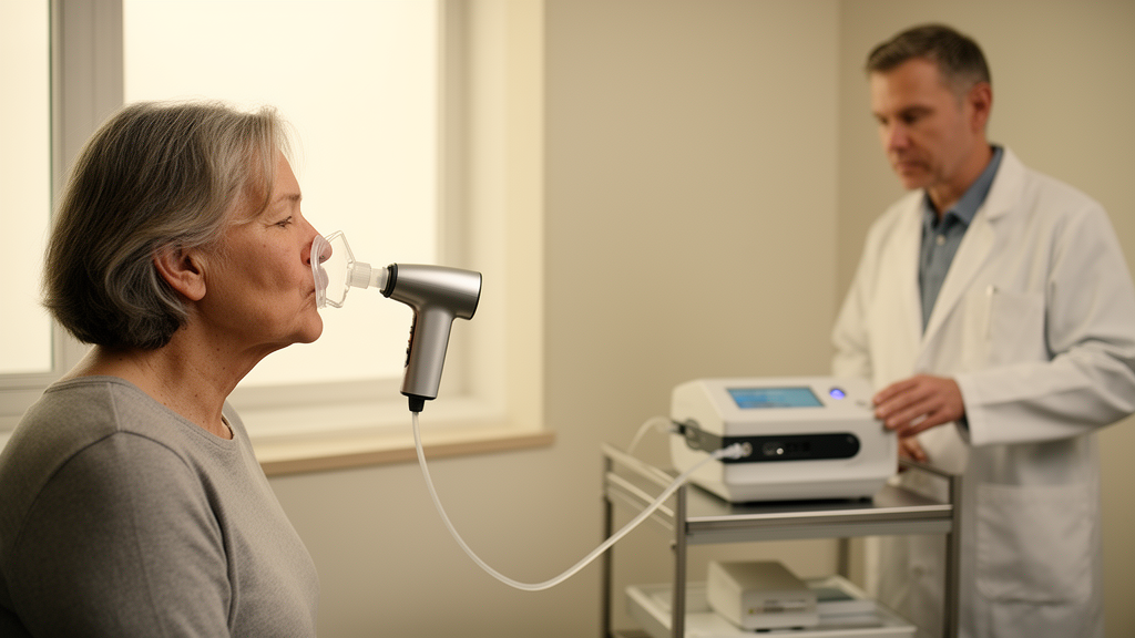
The Griffith study evaluated two complementary technologies operating on very different analytical principles. The first was an electronic nose, or eNose, a device fitted with arrays of chemical sensors that respond to the mixture of volatile compounds in exhaled air. Rather than identifying individual molecules one by one, the eNose captures the overall pattern of breath chemicals, generating a composite profile that machine-learning models can be trained to associate with particular disease states. In this study, those patterns were used to discriminate between breath samples from patients with malignant nodules and those with benign ones.
The second technology was gas chromatography-mass spectrometry, or GC-MS, a gold-standard analytical technique that separates the individual volatile organic compounds in a breath sample and identifies each one by its molecular mass and chemical behavior. Where the eNose reads the forest, GC-MS reads the trees, pinpointing specific compounds that may originate from cancer metabolism. Tumors are known to produce distinctive volatile metabolites, and identifying which of these compounds appear in the breath of patients with malignant nodules offers both diagnostic power and potential biological insight.
Professor Punyadeera reported that both breath-based approaches showed substantially better discrimination between malignant and benign nodules than models built on standard clinical information alone. In practical terms, this means that adding a breath profile to the data clinicians already collect, such as nodule size on CT and patient risk factors, improved the accuracy with which cancerous nodules could be separated from harmless ones. The complementary nature of the two technologies is central to the finding: the pattern-based eNose and the compound-specific GC-MS each contribute independent information, and together they form a more robust diagnostic picture than either could alone.
Importantly, the researchers are careful about what the breath test can and cannot do. Dr Zhang emphasized that while the simple breath test is showing promise in identifying which nodules are most likely to be malignant, it is not intended to replace biopsies. Instead, its role is triage: helping clinicians decide who needs invasive intervention most urgently. Patients whose breath profiles suggest a benign nodule could potentially be spared unnecessary procedures, while those whose profiles flag malignancy could be prioritized for definitive testing. The team hopes the non-invasive test could eventually be incorporated into routine clinical care, reducing the burden of unnecessary interventions for patients with benign nodules.
The implications extend beyond individual patients. A validated breath test is fast, repeatable, and essentially risk-free, making it well suited to monitoring nodules over time and to triaging the growing volume of findings generated by lung cancer screening programs worldwide. The paper, titled Complementary Breath Profiling Using eNose and Gas Chromatography-Mass Spectrometry for Noninvasive Triage of Indeterminate Pulmonary Nodules, was published in MedComm under DOI 10.1002/mco2.71012. As breath research moves from the laboratory toward the clinic, the air we exhale, once considered little more than waste, is emerging as a rich diagnostic medium, and this study offers a concrete step toward a future in which lung cancer triage could begin with nothing more than a breath.
Subject of Research: Breath-based profiling with eNose and GC-MS for non-invasive diagnosis of lung cancer nodules
Article Title: Can your breath help diagnose lung cancer?
Article References: Can your breath help diagnose lung cancer?. Original publication (Bibliographic details unavailable.)
Image Credits: AI Generated
DOI: Not provided
Keywords: lung cancer, breath analysis, electronic nose, gas chromatography-mass spectrometry, pulmonary nodules, non-invasive diagnostics, volatile organic compounds, biopsy, low-dose CT, Griffith University, MedComm, early detection
News Source: Nathaniel Bowman. (October 19, 2026). Breath Test Shows Promise for Distinguishing Cancerous Lung Nodules Without Invasive Biopsies. Scienmag.