Every year, hundreds of thousands of people around the world face the terrifying prospect of losing a foot or a leg to peripheral artery disease, a condition in which narrowed arteries starve the lower limbs of oxygen-rich blood. For surgeons, the standard response is revascularization: reopening or bypassing the blocked vessels to restore flow. Yet the operation does not succeed equally for everyone. A new study from the Netherlands, published in BMC Medical Imaging, has used a glowing fluorescent dye and near-infrared cameras to watch, in real time, how blood actually returns to the feet of patients after such surgery, and the results expose a stark divide between patients with diabetes and those without.
The research, led by Stefan Koning and colleagues at Leiden University Medical Center together with collaborators in The Hague and Copenhagen, enrolled 128 patients with peripheral artery disease, covering 144 treated limbs. Of these, 42 patients, or roughly a third, had diabetes mellitus, accounting for 48 limbs. The team performed quantitative indocyanine green near-infrared fluorescence imaging both before and after revascularization, measuring three time-based perfusion parameters that describe how quickly the fluorescent tracer arrives in the tissue of the foot, how fast it leaves, and how the signal builds up over time. In a subset of 67 patients suffering from chronic limb-threatening ischaemia, the most severe form of the disease, the researchers then checked whether the imaging findings predicted clinical improvement at six weeks.
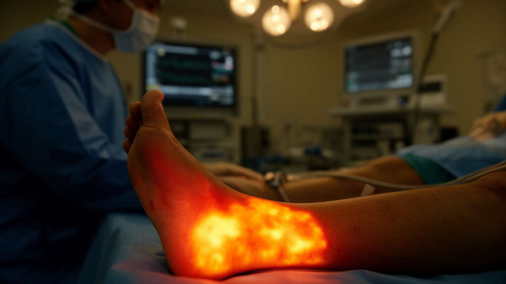
Indocyanine green is a well-established fluorescent tracer that binds to plasma proteins and circulates with the blood. When illuminated by a near-infrared light source, it emits fluorescence that can be captured by specialized cameras, allowing clinicians to visualize perfusion beneath the skin surface without ionizing radiation. Because the technique is dynamic, it does not merely show whether blood vessels are open, as an angiogram does, but whether blood is actually reaching and washing through the microcirculation of the foot, the capillary beds where oxygen delivery ultimately matters. This distinction is crucial, because a technically successful operation on a large artery does not guarantee that the smallest vessels of a diabetic foot can respond.
The preoperative findings were already revealing. Before any intervention, patients with diabetes showed faster inflow and outflow of the tracer than patients without diabetes at most levels of measurement, with the notable exception of the crural level, the region of the lower leg where the arteries branch toward the ankle and foot. This counterintuitive observation suggests that the diabetic foot microcirculation behaves differently even at baseline, a phenomenon the authors link to the complex interplay of neuropathy, microvascular dysfunction and calcified, often rigid vessels that characterize diabetic vascular disease. Rather than simply being more blocked, the diabetic circulation appears to be differently regulated, which may explain why conventional anatomical assessments so often fail to predict outcomes in this group.
The postoperative picture was where the divide became dramatic. After revascularization, only two of the nine perfusion parameters improved significantly in the diabetes group, with p-values below 0.001, whereas six of nine parameters improved in the non-diabetes group, with p-values ranging from 0.027 to below 0.001. In plain terms, opening the arteries produced a far richer physiological response in patients without diabetes. Their feet flushed with restored blood flow across nearly every measurable dimension, while the diabetic feet showed a muted, partial recovery. The imaging was, in effect, capturing the difference between a circulation that can respond to restored supply and one whose microvascular machinery has been damaged by years of metabolic disease.
The clinical correlation strengthened the case. Among the 67 patients with chronic limb-threatening ischaemia, 50 experienced clinical improvement at six weeks, 34 without diabetes and 16 with diabetes, while 14 did not improve, including 8 with diabetes and 6 without, and 3 were lost to follow-up. In the patients who did improve, perfusion rose significantly across all measured parameters in the non-diabetic group, with p-values from 0.08 to below 0.001, but not in the diabetic group. Overall, 85 percent of non-diabetic chronic limb-threatening ischaemia patients improved clinically, compared with 66 percent of diabetic patients, a trend that approached but did not reach statistical significance with a p-value of 0.073. The numbers are small, and the authors are careful to describe the difference as a trend, but the direction is consistent with everything the perfusion imaging showed.
These findings matter because current decision-making in limb-threatening ischaemia relies heavily on the Wound, Ischemia and foot Infection classification system maintained by the Society for Vascular Surgery, which grades the severity of wounds, the degree of ischemia and the presence of infection to predict the likelihood of limb salvage. What that system cannot do is tell a surgeon whether an individual diabetic foot, once reperfused, has enough residual microvascular capacity to heal. The Leiden study suggests that quantitative near-infrared fluorescence imaging could fill that gap, offering a functional, dynamic readout of tissue-level perfusion both before the operation, to set expectations, and immediately after, to verify whether the revascularization has actually changed anything at the capillary level.
The implications for personalized treatment are considerable. If diabetic patients demonstrate reduced perfusion responsiveness after technically successful revascularization, surgeons might use that information to plan adjunctive therapies, to select different revascularization targets, to intensify wound care and offloading, or to have earlier and more honest conversations about prognosis and the risk of amputation. Conversely, a diabetic patient whose postoperative imaging shows a robust perfusion response might be reassured and followed less intensively. The technology itself is relatively practical: indocyanine green is already approved and widely used, the imaging sessions are quick, and the radiation-free nature of near-infrared imaging makes repeated measurements feasible, even at the bedside.
The study, approved by the Medical Research Ethics Committee of Leiden University Medical Center under number G21.087 and conducted in accordance with the Declaration of Helsinki, was co-funded through the Dutch public-private partnership program Health~Holland under the RULER and IMPULSE projects, with additional support from the European Union’s Horizon Europe program through the RETINA project. The work, published open access on 9 September 2026, arrives amid growing recognition that diabetes transforms peripheral artery disease from a plumbing problem into a disease of the smallest vessels and the nerves that regulate them. Diabetic patients face higher rates of neuropathy, more extensive ischemia and dramatically elevated amputation risk, and the study provides direct visual evidence of why.
For now, the authors position their results as a step toward tailored strategies for the highest-risk group rather than a finished clinical tool. The message, however, is unmistakable: restoring flow through a large artery is only half the battle. What ultimately saves a limb is blood arriving where it is needed, in the skin and tissue of the foot, and for the first time surgeons can watch that happen, in glowing green, seconds after injecting a tracer. As near-infrared fluorescence imaging matures, the day may come when every revascularization is followed by a real-time perfusion check, turning an invisible microvascular failure into something a surgeon can see, measure and act upon before the wound, or the limb, is lost.
Subject of Research: Quantitative ICG near-infrared fluorescence imaging of foot perfusion before and after revascularization in peripheral artery disease patients with and without diabetes mellitus
Article Title: Assessment of foot perfusion before and after revascularization using quantitative ICG near-infrared fluorescence imaging in patients with and without diabetes mellitus
Article References: Koning, S., Peul, R. C., Kruiswijk, M. W., Tange, F. P., van Schaik, J., Schepers, A., Brouwers, J. J. W. M., van Rijswijk, C. S. P., van der Bogt, K. E. A., Eiberg, J. P., Vahrmeijer, A. L., Hamming, J. F., van den Hoven, P., & van der Vorst, J. R. (2026). Assessment of foot perfusion before and after revascularization using quantitative ICG near-infrared fluorescence imaging in patients with and without diabetes mellitus. BMC Medical Imaging. https://doi.org/10.1186/s12880-026-02682-9
Image Credits: AI Generated
DOI: 10.1186/s12880-026-02682-9
Keywords: peripheral artery disease, diabetes mellitus, indocyanine green, near-infrared fluorescence imaging, foot perfusion, revascularization, chronic limb-threatening ischaemia, microcirculation, vascular surgery, WIfI classification, amputation risk, BMC Medical Imaging
News Source: Ophelia Keating. (October 18, 2026). Glowing Dye Reveals Why Diabetic Feet Respond Worse to Leg Artery Surgery. Scienmag.