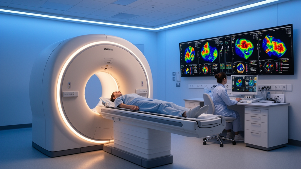
A next-generation total-body PET/CT scanner can produce diagnostic-quality images in as little as one minute of scanning, according to research presented at the 39th Annual Congress of the European Association of Nuclear Medicine (EANM’26) in Vienna. Remarkably, even the fastest images from the new system were rated more highly on average than those produced by a standard PET/CT scanner, suggesting that the technology could fundamentally reshape how clinical imaging is performed around the world.
PET/CT, which combines positron emission tomography with computed tomography, is widely used to diagnose, stage and monitor cancer. Conventional PET/CT scanners have a short axial field of view, meaning they image the body section by section, typically requiring five to six bed positions to cover the body from head to thigh. Long-axial field-of-view (LA-FOV) systems capture a much larger portion of the body at once and detect more of the signal emitted by the radiotracer, raising the possibility of much shorter scans without sacrificing image quality.
In the EQUIP study, researchers at the Peter MacCallum Cancer Centre in Melbourne, Australia, evaluated a next-generation 128 cm LA-FOV PET/CT system, the GE HealthCare Omni 128 cm. Twenty-four participants who had received one of five radiotracers underwent a standard-of-care scan on a conventional short-axial field-of-view (SA-FOV) PET/CT system, followed by a ten-minute scan on the LA-FOV system. The researchers then reconstructed images from the LA-FOV scan using just five, three, two and one minute of data.
Two nuclear medicine specialists, unaware of the acquisition times, graded the images on a five-point scale for overall diagnostic quality, sharpness, lesion conspicuity and image noise. Mean image scores were in the good or excellent range at every acquisition time. Average diagnostic quality scores were 5.0 for the ten- and five-minute images, 4.9 for the three- and two-minute images and 4.5 for the one-minute images. By comparison, the conventional scanner scored 3.9 for diagnostic quality, with lower average scores than the LA-FOV system across all assessed categories and acquisition times.
Lead author Dr Cameron Pain, a nuclear medicine physicist at Peter MacCallum, explained the striking nature of the result. A one-minute scan on this system gave images that readers rated more highly than the standard-of-care scans performed every day. Even when the acquisition time was cut by 90 percent, image quality exceeded current standard-of-care methods.
All images acquired for two minutes or longer scored at least four out of five in every category, and all one-minute images scored at least three out of five. Image scores declined as scan times shortened, with the largest decrease at one minute, particularly for image noise. Even so, these one-minute images were closer in quality to the full ten-minute LA-FOV images than to the standard-of-care SA-FOV images. Objective measurements supported the reader findings, with signal-to-noise ratios in healthy liver tissue remaining broadly consistent between the ten-minute and one-minute images for the radiotracers assessed.
Shorter scans have the potential to make PET/CT more comfortable for patients, particularly those who are in pain or find it difficult to lie still, while also reducing image blurring caused by movement. They could also help imaging departments scan more patients each day. Alternatively, the higher sensitivity of LA-FOV systems could be used to reduce the amount of radiotracer administered, lowering the radiation dose patients receive.
Total-body PET gives choices that have not existed before, Dr Pain noted. Its sensitivity can be used to scan patients faster, to lower the radiation dose and to see disease more clearly. Further research will be needed to confirm these findings. Image quality was assessed using reader scores rather than measures of diagnostic accuracy, so larger studies will be important to confirm that very short scans do not miss small or faint lesions and to determine how faster scanning affects workflow and patient experience in routine clinical practice.
The EQUIP findings arrive as long-axial field-of-view total-body PET systems gain regulatory approvals and enter clinical service more broadly. If subsequent studies confirm that one-minute acquisitions preserve diagnostic accuracy, the change could ripple through oncology departments worldwide, easing burdens on patients who struggle with long scans while expanding daily throughput. For a technology long constrained by the physics of counting photons, the ability to compress a scan from ten minutes to one, while exceeding the quality of today’s standard of care, marks a striking demonstration of what next-generation detector sensitivity can deliver.
The physics behind this gain in speed lies in geometric efficiency. In a conventional scanner, a radiotracer signal is only counted when the pair of annihilation photons strikes detectors on opposite sides of a narrow ring, so most photons emitted by the patient escape undetected. A scanner whose detector ring spans roughly the entire torso captures a far larger fraction of that emission, and the sensitivity advantage scales dramatically — often by a factor of several dozen compared with short-axial systems. That surplus of detected signal is what allows reconstruction algorithms to form a usable image from a fraction of the usual counting statistics, and it explains why the one-minute images in the EQUIP study could still compete with, and exceed, conventional standard-of-care acquisitions lasting far longer.
The five radiotracers used in the study also illustrate how versatile such a platform could be. Fluorine-18 FDG remains the workhorse of oncological PET, mapping glucose metabolism across a broad range of cancers. Prostate-specific membrane membrane tracers such as fluorine-18 DCFPyL and gallium-68 PSMA agents target prostate cancer with high specificity, while gallium-68 DOTATATE images neuroendocrine tumours through somatostatin receptors. Iodine-124, a longer-lived positron emitter, serves niche applications where its half-life permits imaging days after injection. Demonstrating consistent image quality across this pharmacological spectrum matters because a scanner’s sensitivity dividend is only clinically meaningful if it holds regardless of which tracer a department deploys.
Reduced acquisition time also interacts with patient physiology in ways that go beyond comfort. PET images are inherently vulnerable to motion: a patient breathing, shifting position, or simply unable to remain still over a five-to-six-bed-position protocol introduces blur that can obscure small lesions. Compressing the acquisition into a single minute narrows the window during which motion can degrade the data, which is particularly relevant for imaging the lungs, liver and other structures that move with respiration. For patients in pain, children, or those with claustrophobia or cognitive impairment, the difference between a tolerable and an intolerable examination can determine whether staging information is obtained at all.
The throughput implications deserve attention as well. Nuclear medicine departments in many health systems face backlogs driven by limited scanner availability and the sequential nature of conventional protocols, in which each patient occupies the table for the full duration of a multi-bed acquisition. If diagnostic-quality images can be obtained in a minute or two, the number of patients scanned per day could rise substantially, or alternatively the spare sensitivity could be traded for lower injected activity — reducing radiation exposure, easing constraints on radiopharmacy production, and potentially making scans feasible for populations, such as pregnant patients or those requiring repeated surveillance imaging, in whom dose is a particular concern.
Objective metrics reinforce the subjective reader scores. Signal-to-noise ratio measured in healthy liver tissue remained broadly consistent between the ten-minute and one-minute reconstructions for the tracers assessed, indicating that the statistical penalty of a shorter acquisition was largely offset by the scanner’s sensitivity rather than by aggressive smoothing or noise-suppression techniques that can mask genuine pathology. This distinction is important, because an image that looks clean but suppresses subtle uptake could mislead clinicians even when quantitative measures appear favourable.
Nevertheless, the study’s design leaves important questions open. Reader scores capture perceived quality — sharpness, conspicuity, noise — but not whether a scan changes a diagnosis, upstages a tumour, or detects a metastasis that would otherwise be missed. Sensitivity analyses powered on clinical endpoints, involving larger and more heterogeneous cohorts, will be needed before one-minute protocols can be adopted with confidence, particularly for small or faint lesions where the one-minute images showed their greatest degradation in noise. How such protocols integrate with CT components of the examination, radiotracer uptake waiting times, and reporting workflows will also shape whether the theoretical throughput gains materialise in practice.
The EQUIP study is registered as a formal clinical trial, reflecting a deliberate effort to move total-body PET from a research curiosity toward routine clinical tool. As long-axial systems receive regulatory clearances and proliferate beyond a handful of pioneering centres, the central question shifts from whether the hardware works to how its extraordinary sensitivity should best be spent — on speed, on dose reduction, or on image clarity. The Melbourne findings suggest these need not be mutually exclusive, and that the trade-offs clinicians have long accepted as inherent to PET may be far more negotiable than previously assumed.
Subject of Research: Total-body PET imaging
Article Title: Next-generation total-body PET scanner can produce diagnostic-quality images in as little as one minute
Article References: Next-generation total-body PET scanner can produce diagnostic-quality images in as little as one minute. Original publication (Bibliographic details unavailable.)
Image Credits: AI Generated
DOI: Not provided
Keywords: Next-generation, total-body, scanner, produce, diagnostic-quality, images, little, minute, scientific research
News Source: Ophelia Keating. (October 18, 2026). One-minute total-body PET scans deliver diagnostic-quality images in EQUIP study. Scienmag.