Every year, acute lymphoblastic leukemia claims thousands of lives, many of them children, and the first clue to its presence often hides in a single drop of blood viewed under a microscope. Now researchers at Vellore Institute of Technology in India have built an artificial intelligence system that can distinguish leukemic cells from healthy ones with 99 percent accuracy, and the way it does so is as fascinating as the result itself. The new model, called MoViTHAFX-Net, doesn’t rely on a single neural network design. Instead, it fuses two fundamentally different ways of seeing into one hybrid brain, combining the fine-grained eye of a convolutional network with the panoramic gaze of a transformer.
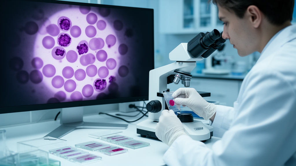
The study, published in Scientific Reports, tackles a deceptively simple question: can a computer reliably tell an acute lymphoblastic leukemia blast apart from a healthy hematogone in a peripheral blood smear image? The answer matters because microscopic screening remains one of the most accessible diagnostic tools in oncology, yet it depends entirely on human expertise and human attention. A pathologist scanning hundreds of cells can miss subtle morphological differences, and fatigue, workload, and experience all shape what gets noticed. An automated assistant that flags suspicious cells could support that screening process, catching patterns that even trained eyes might overlook.
What makes MoViTHAFX-Net unusual is its dual architecture. On one side sits MobileNet, a lightweight convolutional neural network originally designed for mobile devices, which excels at extracting local morphological information. It examines the fine details of a cell: the texture of the nucleus, the shape of the membrane, the granularity of the cytoplasm. On the other side sits a Vision Transformer, or ViT, a model that treats an image as a sequence of patches and models long-range contextual relationships between them. Where MobileNet sees the tree, the ViT sees the forest. It understands how distant regions of a cell relate to each other, capturing the global structure that defines a leukemic blast.
But simply running two networks in parallel and gluing their outputs together isn’t enough. The researchers introduced a component called Hierarchical Attention Fusion, or HAF, which first refines the features from both branches and then adaptively combines three learnable morphology-guided feature projections before the final binary classification. In plain terms, HAF doesn’t just merge the two streams of information; it decides, cell by cell, how much weight to give each type of feature. A blast with an unusual nuclear shape might trigger stronger attention to the MobileNet branch, while a cell whose overall structure is telling might shift focus to the transformer. This adaptive weighting is what gives the hybrid model its edge over a direct concatenation of the two networks, as the ablation results in the study demonstrate.
The training data came from the C-NMC 2019 dataset, a benchmark collection of 10,661 labelled cell images from peripheral blood smears. The researchers used an image-level stratified partition, splitting the data into 7462 training images, 1600 validation images, and 1599 held-out test images. Data augmentation, which artificially expands the training set by applying transformations like rotations and flips, was restricted to the training partition to keep the validation and test sets clean. This careful separation ensures the model isn’t accidentally memorizing images it has already seen, a common pitfall in machine learning studies.
On the held-out test partition, the model correctly classified 1583 of 1599 images, corresponding to 99.00 percent accuracy. The precision, recall, and F1-score all came in at approximately 0.990, and the area under the curve, a measure of the model’s ability to distinguish between classes across all possible thresholds, reached 0.99. These numbers mean the model almost never mistakes a healthy cell for a leukemic one, and almost never lets a leukemic cell slip past undetected. In diagnostic terms, that balance between false alarms and missed detections is exactly what a screening tool needs.
Yet the researchers are careful about what these numbers actually prove. Because the reported custom split is image-level rather than subject-disjoint, and evaluation is limited to a single benchmark dataset, the results should be interpreted as internal benchmark performance rather than evidence of patient-independent or multicentre clinical generalization. In other words, the model excels at the specific dataset it was trained on, but whether it would perform equally well on blood smears from different hospitals, different staining protocols, or different patient populations remains an open question. This kind of transparent reporting is rare and valuable in a field where inflated claims often outpace clinical reality.
The team also added a qualitative layer to the system: Grad-CAM++ visualization, a post hoc technique that highlights the image regions contributing to each prediction. When the model flags a cell as a leukemic blast, Grad-CAM++ generates a heatmap showing which parts of the image drove that decision, typically the nucleus or other morphologically significant regions. This transparency is crucial for building trust with pathologists, who need to understand why an AI system reached a conclusion before they act on it. It transforms the model from a black box into something closer to a collaborative partner that shows its work.
The implications reach beyond leukemia. The hybrid CNN-transformer approach with adaptive attention fusion could apply to any classification problem where both local detail and global structure matter, from detecting tumors in histopathology slides to identifying defects in industrial imaging. The architecture demonstrates that combining complementary neural network designs, rather than betting on a single paradigm, can yield measurable gains. As ablation results within the same experimental setting indicate an incremental advantage of Hierarchical Attention Fusion over direct CNN-ViT concatenation, the study offers a concrete design lesson for future hybrid models.
For now, MoViTHAFX-Net remains a research tool, not a clinical one. But the trajectory is clear: as hybrid architectures mature and datasets grow more diverse, models like this could move from benchmark datasets to real diagnostic workflows, supporting microscopic screening in laboratories around the world. The study, published open access, received no external funding and was conducted by A. Elavarasi and N. Jaisankar of the School of Computer Science and Engineering at Vellore Institute of Technology. It represents a step toward a future where the first line of defense against leukemia isn’t a tired human eye, but a tireless hybrid intelligence that sees both the tree and the forest.
Subject of Research: Deep hybrid MobileNet and vision transformer model for acute lymphoblastic leukemia classification from blood smear images.
Article Title: A deep hybrid feature extraction model based on MobileNet and vision transformer for acute lymphoblastic leukemia prediction
Article References: Elavarasi, A., & Jaisankar, N. (2026). A deep hybrid feature extraction model based on MobileNet and vision transformer for acute lymphoblastic leukemia prediction. Scientific Reports. https://doi.org/10.1038/s41598-026-73064-y
Image Credits: AI Generated
DOI: 10.1038/s41598-026-73064-y
Keywords: acute lymphoblastic leukemia, MobileNet, vision transformer, deep learning, C-NMC 2019, Grad-CAM++, hierarchical attention fusion, medical imaging, blood smear, machine learning, cancer detection, deep
News Source: Nathaniel Bowman. (October 17, 2026). Hybrid AI Spots Leukemia Cells With 99% Accuracy in Blood Smears. Scienmag.