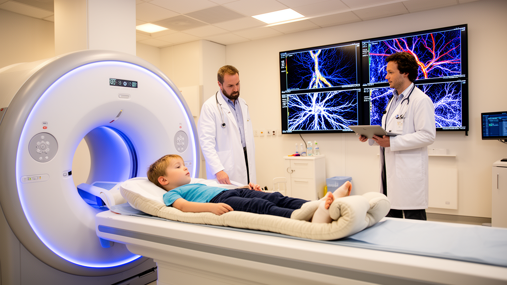
For decades, the most sensitive non-invasive window into the inflamed joints of children with juvenile idiopathic arthritis has been magnetic resonance imaging. Yet conventional MRI protocols have always had a blind spot: they can show the swollen, contrast-enhancing synovial membrane lining the joint, but they cannot simultaneously reveal the vascular changes that accompany the inflammation. A new study published in Pediatric Radiology by a team at the Children’s Hospital of Fudan University in Shanghai now closes that gap. Using an ultra-high-field 5.0-Tesla scanner together with contrast-enhanced magnetic resonance angiography, the researchers captured, in a single examination, both the synovial inflammation and the arterial and microcirculatory alterations in the hands of children with JIA — a combined view they describe as the synovium-vascular axis.
The technical significance of the 5.0-T platform deserves emphasis. Clinical MRI has long been dominated by 1.5-T and 3-T scanners, and the jump to 5.0-T brings a substantially higher signal-to-noise ratio, which can be traded for finer spatial resolution, faster acquisition, or both. For small structures such as the finger joints and the palmar digital arteries of children, that resolution matters enormously. The synovium of a healthy child is less than a millimeter thick, and detecting pathological thickening, enhancement, and the fine vascular architecture of the hand demands a field strength that can resolve millimeter-scale anatomy with confidence. The study, led by Qiong Yao and Guanke Cai as co-first authors under the corresponding authorship of Hao Li and Xihong Hu, optimized dedicated 5.0-T MRI and CE-MRA protocols for pediatric hand imaging, with Kai Lu of United Imaging Research contributing to protocol development and Xin Guo handling vascular image post-processing.
The design was straightforward but rigorous. Twenty children with JIA and ten age- and sex-matched healthy controls underwent hand MRI at 5.0-T, including contrast-enhanced T1-weighted imaging and CE-MRA. From these images the team quantified a battery of parameters spanning both sides of the synovium-vascular axis: maximum synovial thickness, the synovial enhancement rate, a bone marrow edema score, the diameter ratios of the common palmar digital arteries, palmar arch integrity, a distal microcirculation score, and vascular tortuosity. These imaging markers were then correlated with clinical disease activity measures — the Juvenile Arthritis Disease Activity Score-27 (JADAS-27), the erythrocyte sedimentation rate, and C-reactive protein — the standard laboratory and clinical yardsticks used in pediatric rheumatology.
The results paint a strikingly coherent picture of what happens to the inflamed child’s hand. Compared with controls, the JIA group showed dramatically higher maximum synovial thickness, at 5.19 plus or minus 2.35 millimeters versus 0.83 plus or minus 0.24 millimeters in healthy children, a difference significant at P less than 0.001. The synovial enhancement rate was more than doubled, at 0.44 versus 0.20, and the median bone marrow edema score was 7.50 in the JIA group compared with zero in controls. In other words, the 5.0-T scanner could resolve synovial pathology at a scale that clearly separated diseased from healthy tissue, while simultaneously documenting the bone marrow inflammation that is a known harbinger of erosive joint damage.
But the vascular findings are what elevate this study beyond a conventional MRI assessment. The common palmar digital artery diameter ratio was significantly lower in the JIA group, at 0.690 versus 1.000 in controls, indicating measurable narrowing of the arteries supplying the fingers. The distal microcirculation score was markedly reduced, at a median of 1.0 versus 3.0, showing impaired perfusion at the fingertips. And vascular tortuosity — a measure of abnormally twisted vessels — was increased, with a median of 2.0 versus zero. These are precisely the changes one would predict from the biology of chronic synovial inflammation, in which angiogenesis, hypoxia, and remodeling of the periarticular vasculature are known features of inflammatory arthritis, but which have never before been quantified in children alongside synovial metrics in a single 5.0-T examination.
The correlations with clinical disease activity tie the imaging findings to the real-world management of JIA. The bone marrow edema score correlated positively with JADAS-27 at r equal to 0.674, the maximum synovial thickness at r equal to 0.581, and the enhancement rate at r equal to 0.634, all statistically significant. In a regression analysis, the bone marrow edema score was independently associated with the logarithm of JADAS-27, with a standardized beta of 0.677 and P equal to 0.005. This means that the single strongest imaging predictor of overall disease activity was not the visible synovial swelling but the invisible bone marrow inflammation — a finding consistent with the adult rheumatoid arthritis literature, where bone marrow edema on MRI is recognized as a predictor of erosive progression and a marker of active disease.
What makes this work genuinely novel is the demonstration that synovial inflammation, arterial narrowing, and impaired distal microcirculatory perfusion form a distinct, quantifiable pattern in children with JIA. The concept of a synovium-vascular axis — the idea that the inflamed synovium and the periarticular vasculature are two faces of the same pathological process — has been discussed in the adult inflammatory arthritis literature for years, but it has never been mapped in children with this combination of ultra-high-field resolution and dedicated vascular angiography. The CE-MRA component, in particular, allows the palmar arch, the digital arteries, and the distal microcirculation to be assessed non-invasively, something previously possible only with invasive catheter angiography or, indirectly, with ultrasound Doppler techniques that cannot visualize the deep palmar arch reliably.
The clinical implications are considerable. If the vascular parameters documented here — arterial diameter ratios, microcirculation scores, and tortuosity — can be validated in larger cohorts, they could become exploratory imaging biomarkers that complement JADAS-27 and laboratory markers in assessing disease activity and treatment response. This would be particularly valuable in children, in whom laboratory markers such as ESR and CRP can be normal even during active disease, and in whom repeated exposure to ionizing radiation from conventional angiography or computed tomography is undesirable. MRI, by contrast, involves no ionizing radiation, and the 5.0-T platform’s higher signal-to-noise ratio could allow shorter scan times — an important consideration for pediatric patients who must lie still for extended periods.
The authors are careful to frame their vascular parameters as candidate exploratory imaging markers requiring validation in larger multicenter cohorts, and this caution is warranted. The study involved twenty children and ten controls from a single center, and the correlations, while statistically robust, need replication before they can inform clinical decisions. Nevertheless, the demonstration that a single 5.0-T examination can simultaneously quantify synovial inflammation, bone marrow edema, arterial narrowing, and microcirculatory impairment represents a meaningful advance in pediatric imaging. It suggests that the synovium-vascular axis is not merely a conceptual framework but a measurable clinical reality in children with JIA, and it opens the door to a more comprehensive, radiation-free assessment of the disease that could ultimately change how pediatric rheumatologists monitor and treat their youngest patients.
Subject of Research: Health
Article Title: 5.0-T magnetic resonance imaging and contrast-enhanced magnetic resonance angiography for the evaluation of juvenile idiopathic arthritis
Article References: Yao, Q., Cai, G., Yang, L., Shen, J., Liu, H., Sun, L., Lu, K., Guo, X., Li, H., & Hu, X. (2026). 5.0-T magnetic resonance imaging and contrast-enhanced magnetic resonance angiography for the evaluation of juvenile idiopathic arthritis. Pediatric Radiology. https://doi.org/10.1007/s00247-026-06809-6
Image Credits: AI Generated
DOI: 10.1007/s00247-026-06809-6
Keywords: juvenile idiopathic arthritis, MRI, magnetic resonance angiography, pediatric radiology, 5.0-T MRI, microcirculation, synovitis, bone marrow edema, JADAS-27, CE-MRA, magnetic, resonance
News Source: Nathaniel Bowman. (October 17, 2026). 5-Tesla MRI Reveals Hidden Vascular Damage in Children With Juvenile Arthritis. Scienmag.