A recent letter to the editor published in the Journal of Cellular and Molecular Medicine has ignited a sharp debate within the cerebrovascular research community, challenging the interpretation of a high-profile transcriptomic study on Moyamoya disease. The original investigation, led by Chen and colleagues, applied transcriptome-wide RNA sequencing to temporal muscle tissue harvested during indirect revascularization surgery, uncovering a striking association between collagen-rich extracellular matrix remodelling and the formation of postoperative collateral vessels. The letter, authored by Emaan Rani and Mirza Faaz Baig, acknowledges the creativity and molecular ambition of the study but raises fundamental concerns about cohort design, surgical comparators, and clinical phenotyping that could reshape how the field approaches surgical genomics in this rare and devastating cerebrovascular disorder.
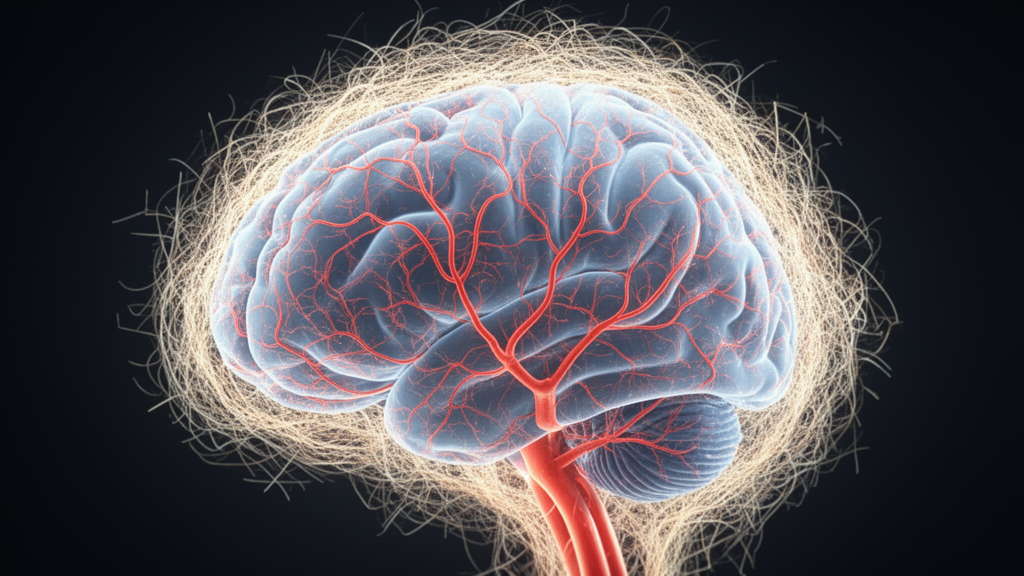
Moyamoya disease is a chronic, progressive occlusion of the terminal internal carotid arteries and their proximal branches, characterised by the development of a fragile network of compensatory collateral vessels at the base of the brain that resembles a puff of smoke on angiographic imaging. When medical therapy alone cannot prevent recurrent ischemic or hemorrhagic events, surgeons turn to revascularization procedures. Indirect bypass, the technique central to the Chen study, involves laying vascularised tissue such as the temporalis muscle directly onto the cortical surface, relying on the patient’s own biological capacity to sprout new arteries from the donor tissue into the ischemic brain over weeks to months. The molecular signals that govern this neoangiogenesis have remained poorly characterised, which is precisely why the transcriptomic profiling approach attracted so much attention.
The Chen team’s central finding was that genes associated with extracellular matrix organisation and collagen deposition were significantly enriched in patients who went on to develop robust collateral circulation after surgery, as graded by the Matsushima angiographic scale. This suggested that the structural scaffold laid down by the transplanted temporal muscle might serve as a permissive microenvironment through which endothelial cells migrate, proliferate, and anastomose with the cortical vasculature. The implication was tantalising: if collagen-rich matrix remodelling is a driver rather than a byproduct of successful collateralisation, then pharmacological modulation of matrix biology could one day enhance surgical outcomes in patients whose native angiogenic response is insufficient.
However, Rani and Baig argue that this conclusion rests on a cohort whose internal heterogeneity was never adequately controlled. Their first and most pointed criticism concerns the pooling of paediatric and adult patients without stratification by clinical disease stage, whether measured by the Suzuki angiographic staging system or by hemodynamic assessment of cerebral perfusion reserve. Children with Moyamoya disease possess a dramatically higher native capacity for neoangiogenesis and collateral development than adults, a biological trait that operates independently of any measurable difference in extracellular matrix gene expression. When these two age groups are analysed together, the resulting transcriptomic signature may simply reflect an age-specific baseline angiogenic response rather than a shared molecular mechanism of surgical success.
The letter elaborates on this confounding problem with technical precision. Endogenously generated differential angiogenic signals are themselves associated with advanced disease stage and severe basal hypoperfusion, meaning that the ischemic drive within the brain tissue of a stage IV patient is fundamentally different from that of a stage II patient. Without correcting multi-variable transcriptomic models for pre-operative Suzuki staging and cerebrovascular reserve capacity, the collagen-rich signature identified by Chen and colleagues could be an artifact of mixed disease severity rather than a genuine molecular predictor of collateral formation. Rani and Baig point to prior work by Bao and colleagues, published in Frontiers in Neurology, which demonstrated that the efficacy of indirect revascularization is intrinsic to an exquisite balance between disease stage progression and age-associated neoangiogenic potential, with different patient subsets exhibiting markedly different collateral formation trajectories.
The second major objection concerns the absence of a surgical comparator group. The Chen study included only patients who underwent indirect bypass, with no tissue samples from individuals who received direct bypass procedures, such as superficial temporal artery to middle cerebral artery anastomosis, or combined approaches that pair direct bypass with encephaloduroarteriomyosynangiosis. This distinction is not merely academic. Direct bypass restores hemodynamic function immediately by creating a high-flow surgical connection between extracranial and intracranial circulation, fundamentally altering shear stress and metabolic conditions in the adjacent cortical tissue from the moment the clips are released. Indirect bypass, by contrast, depends entirely on tissue-level angiogenesis unfolding over an extended postoperative period. The molecular programs activated in temporal muscle under these two radically different hemodynamic regimes are unlikely to be identical, yet without comparative tissue from combined or direct bypass patients, the Chen results cannot be generalised across surgical techniques.
Rani and Baig reference a retrospective study by Zhao and colleagues published in Experimental and Therapeutic Medicine, which examined 76 combined direct and indirect bypass procedures in adult Moyamoya patients and found significantly different clinical and angiographic outcomes compared to standalone indirect procedures, along with distinct local tissue perfusion pressures. They argue that the addition of tissue controls from patients undergoing combined direct-indirect bypass would allow future transcriptomic studies to separate expression patterns of extracellular matrix molecules that are specific to the indirect angiogenic process from those that merely reflect general tissue healing under different flow conditions. This is a critical distinction for any researcher hoping to translate collagen biology into a therapeutic target, because a signature that appears in all postoperative temporal muscle regardless of surgical approach is unlikely to be the molecular key to collateral vessel formation.
The third concern addresses clinical phenotyping, or rather the lack thereof. The Chen study grouped patients into binary collateral outcomes using the Matsushima grading system but did not account for the primary clinical presentation, namely whether patients presented with ischemic or hemorrhagic Moyamoya disease. These two phenotypes are not interchangeable variants of the same illness. Ischemic and hemorrhagic Moyamoya differ in their underlying pathophysiological mechanisms, in the degree of vessel wall inflammation, and in the postoperative trajectory of collateral formation. Hemorrhagic Moyamoya is characterised by microvascular fragility and altered expression of matrix metalloproteinases, enzymes that directly degrade extracellular matrix components and are intimately connected to temporal muscle tissue biology and postoperative angiogenesis. The unchecked accumulation of both phenotypes within a single transcriptomic cohort risks blending phenotype-specific molecular drivers of collateralisation into an averaged signal that corresponds to neither group.
Rani and Baig cite work by Ge and colleagues published in BMC Neurology, which specifically examined postoperative collateral formation after indirect bypass in hemorrhagic Moyamoya disease and found that collateral dynamics and angiographic response differ according to aetiology. They call for subgroup analyses of ischemic and hemorrhagic patients to determine whether the collagen-rich extracellular matrix signature identified by Chen and colleagues is preserved across both presentations or whether it is a molecular fingerprint unique to one phenotype. If the signature diverges between ischemic and hemorrhagic Moyamoya, then any future therapeutic strategy aimed at enhancing matrix remodelling would need to be tailored to the specific clinical context, a level of precision that the current mixed-cohort analysis simply cannot support.
In their concluding remarks, Rani and Baig strike a constructive tone, acknowledging that Chen and colleagues have provided a much-needed molecular underpinning of the relationship between extracellular matrix remodelling, collagen deposition in temporal muscle tissue, and successful postoperative collateralisation in Moyamoya disease. They emphasise, however, that angiogenic potential, disease stage, surgical approach variability, and clinical phenotype remain critical variables that must be addressed before these transcriptomic discoveries can be translated into targeted therapeutic tools. Their prescription is clear: multi-centre, phenotype-stratified, prospective cohorts with age-matched paediatric and adult subgroups, corrected for pre-operative Suzuki staging and cerebrovascular reserve, and including tissue comparators from direct and combined bypass procedures. Only through such rigorous design, they argue, can the field determine whether collagen-rich extracellular matrix biology is a genuine driver of collateral vessel formation or a molecular echo of patient heterogeneity.
Subject of Research: Moyamoya disease transcriptomics and postoperative angiogenesis
Article Title: Letter to Editor on Collagen‐Rich Extracellular Matrix and Postoperative Angiogenesis in Moyamoya Disease: The Need for Phenotype‐Stratified Transcriptomic Analysis
Article References: Rani, E., & Baig, M. F. (2026). Letter to Editor on Collagen‐Rich Extracellular Matrix and Postoperative Angiogenesis in Moyamoya Disease: The Need for Phenotype‐Stratified Transcriptomic Analysis. Journal of Cellular and Molecular Medicine, 30(19), Article e71400. https://doi.org/10.1111/jcmm.71400
Image Credits: AI Generated
DOI: 10.1111/jcmm.71400
Keywords: Moyamoya disease, transcriptomics, collagen, extracellular matrix, angiogenesis, revascularization, cerebrovascular, letter to editor, Letter, Editor, Rich, Extracellular
News Source: Brooke Gardner. (October 17, 2026). Collagen-Rich Matrix and Postoperative Angiogenesis in Moyamoya Disease: A Call for Phenotype-Stratified Transcriptomics. Scienmag.