Human fat cells are notoriously fragile laboratory subjects. Mature adipocytes — the large, lipid-filled cells that make up most of our adipose tissue — are buoyant, greasy, and easily torn apart by the shear forces generated when bioinks are pushed through a printing nozzle. Yet these are precisely the cells that researchers need in three-dimensional culture if they want to study obesity, insulin resistance, and type 2 diabetes in a human-relevant setting. A team led by Petra J. Kluger at the University of Stuttgart has now demonstrated a way to do exactly that: a support-bath bioprinting technique that embeds fully differentiated human adipocytes into precisely shaped 3D constructs without destroying their structure or their metabolism. The work, published in Engineering in Life Sciences on 10 August 2026, offers a practical route toward laboratory-grown fat tissue that behaves like the real thing.
The challenge the researchers faced is fundamental to the biology of the cells themselves. Adipocytes store most of their volume as a single large lipid droplet, which makes them less dense than the aqueous media surrounding them. In a liquid or semi-liquid bioink, they float, drift, and cluster rather than staying where the printer places them. Their hydrophobic surfaces and large size — often 50 to 100 micrometers across — mean that standard extrusion through narrow nozzles tends to rupture their membranes. Most existing 3D adipose models sidestep the problem by using adipose-derived stem cells that are coaxed into becoming fat cells after printing. That approach works, but it produces less mature tissue with different signaling behavior. Fully differentiated adipocytes are more physiologically faithful, which is why the Stuttgart team chose to print them directly, despite the mechanical risks.
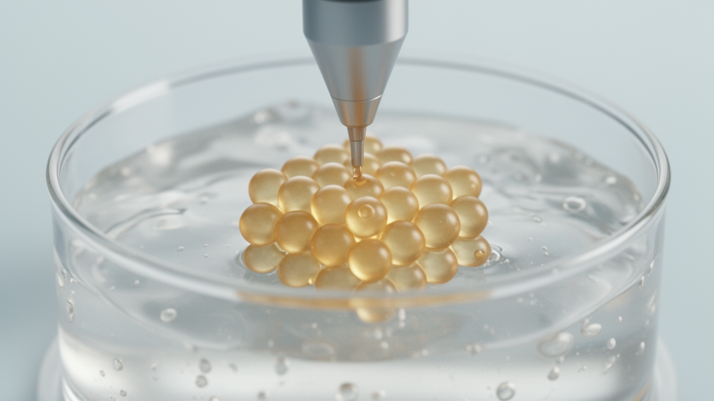
Their solution combines two gels with complementary roles. The cells are suspended in a low-concentration gellan gum bioink, a polysaccharide gel that is fluid enough to be extruded gently but can be stabilized after deposition by adding calcium chloride, which crosslinks the polymer chains. The printed material is not deposited into open air, where a soft bioink would simply slump and spread. Instead, it is extruded into a chilled agarose support bath — a self-standing gel that holds each filament in place the moment it leaves the nozzle. This technique, sometimes called embedded or freeform bioprinting, counteracts both the spreading of the ink and the buoyancy of the cells, allowing accurate layer-by-layer construction of soft structures that would collapse under conventional printing.
To find a formulation that balanced fluidity with stability, the researchers isolated mature adipocytes from human adipose tissue obtained during elective surgery and mixed them into gellan gum at a final polymer concentration of 0.3 percent. They tested formulations containing different concentrations of phosphate-buffered saline and measured the viscosity of both the bioink and a 0.5 percent agarose support bath across a range of shear rates. The goal was a bioink that would flow smoothly under gentle pressure yet hold its shape once crosslinked, and a support bath that would remain solid enough to confine the printed filaments without crushing them. The optimization identified a combination that satisfied both requirements, allowing the team to extrude the cell-laden ink through a 600-micrometer nozzle at pressures of 4 to 6 kilopascals and speeds of 14 to 16 millimeters per second — conditions mild enough to spare the delicate cells.
The structural results were striking. When the same soft bioink was printed without a support bath, it spread after extrusion and could not be layered reliably into defined shapes. With the agarose bath, the team produced stable grid structures measuring 20 by 20 by 1 millimeters with uniform pores and well-aligned filaments. Quantitative comparison against the computer-aided design showed remarkably low deviation: construct length differed by only 2.66 percent, pore width by 2.87 percent, and filament width by 3.35 percent. For a material soft enough to mimic adipose tissue — which is among the softest tissues in the body — that level of geometric fidelity is a meaningful technical achievement, since it demonstrates that printing accuracy and physiological softness need not be traded against each other.
Cell survival was the second critical test. Live and dead staining showed that most adipocytes remained viable through three days after printing, and viable cells were still observed on day eight in an exploratory assessment. One telling indicator came from measurements of lactate dehydrogenase, an enzyme that leaks out of cells when their membranes are damaged. Release dropped significantly from 29.09 percent on day one to 9.66 percent on day three, suggesting that the transient membrane stress caused by printing was real but recoverable — the cells repaired themselves once settled into their new gel environment. This pattern matters because it distinguishes a gentle process that temporarily perturbs cells from a harsh one that permanently destroys them.
Morphology and function held up as well. Lipid staining revealed uniformly distributed, lipid-filled cells throughout the constructs, and expression of perilipin A — a protein that coats lipid droplets and is a standard marker of intact adipocyte structure — confirmed that the cells’ characteristic unilocular architecture had been preserved. Most importantly for anyone hoping to model metabolism, the printed cells still responded to hormonal stimulation. When the researchers applied isoproterenol, a beta-adrenergic agonist that triggers lipolysis, the cells released glycerol in dose-appropriate fashion: from 40.60 to 137.88 nanograms per milliliter on day one, and from 65.86 to 200.54 nanograms per milliliter on day three. In other words, the printed fat tissue could still perform its core metabolic job of breaking down stored lipid on command.
Why does this matter beyond the printing bench? Adipose tissue is not merely passive storage; it is a metabolically active organ involved in energy storage, endocrine signaling, and whole-body metabolic regulation. Dysfunction of adipocytes contributes to obesity, insulin resistance, type 2 diabetes, and other diseases, which creates strong demand for human-relevant laboratory models. Two-dimensional cultures cannot reproduce the soft mechanical environment, spatial organization, or buoyancy effects of native fat tissue, while animal models suffer from species differences as well as ethical and financial constraints. A 3D construct built from genuine mature human adipocytes, held in a defined geometry, closes much of that gap and could support preliminary drug and cosmetic testing before animal or clinical studies.
The Stuttgart team is candid about the limits of the current work. The culture periods examined were short, and real adipose tissue depends on interactions with endothelial cells, immune cells, and stromal cells, as well as on a functioning vasculature that supplies oxygen to cells living deep within tissue. Extending culture duration, introducing vascularization, and incorporating those additional cell types are the obvious next steps. The support-bath method, however, provides a practical foundation on which such complexity can be built, because it solves the hardest problem first: keeping mature adipocytes alive and functional while placing them exactly where a design requires.
The broader significance lies in accessibility. The workflow relies on relatively inexpensive materials — gellan gum and agarose are common, food-grade polysaccharides — and standard extrusion bioprinting equipment, making the approach reproducible across laboratories without exotic hardware. If follow-up studies confirm the method at longer time scales and in more complex, vascularized constructs, printed human fat tissue could become a routine platform for studying metabolic disease, screening compounds that affect fat cell function, and eventually engineering soft tissue for regenerative and reconstructive medicine. For a cell type long considered too fragile to print, the fat cell has just become considerably more approachable.
Subject of Research: Support-bath bioprinting of mature human adipocytes in gellan gum hydrogels for functional 3D adipose tissue models
Article Title: Support-bath bioprinting creates functional 3D human fat cell models
Article References: Support-bath bioprinting creates functional 3D human fat cell models. (n.d.). Original publication
Image Credits: AI Generated
DOI: Not provided
Keywords: bioprinting, adipocytes, support bath, gellan gum, agarose, 3D tissue models, obesity, metabolic disease, adipose tissue, tissue engineering, lipolysis, gellan gum bioink
News Source: Gregory Coleman. (October 17, 2026). Gentle support-bath bioprinting keeps mature human fat cells alive in 3D. Scienmag.