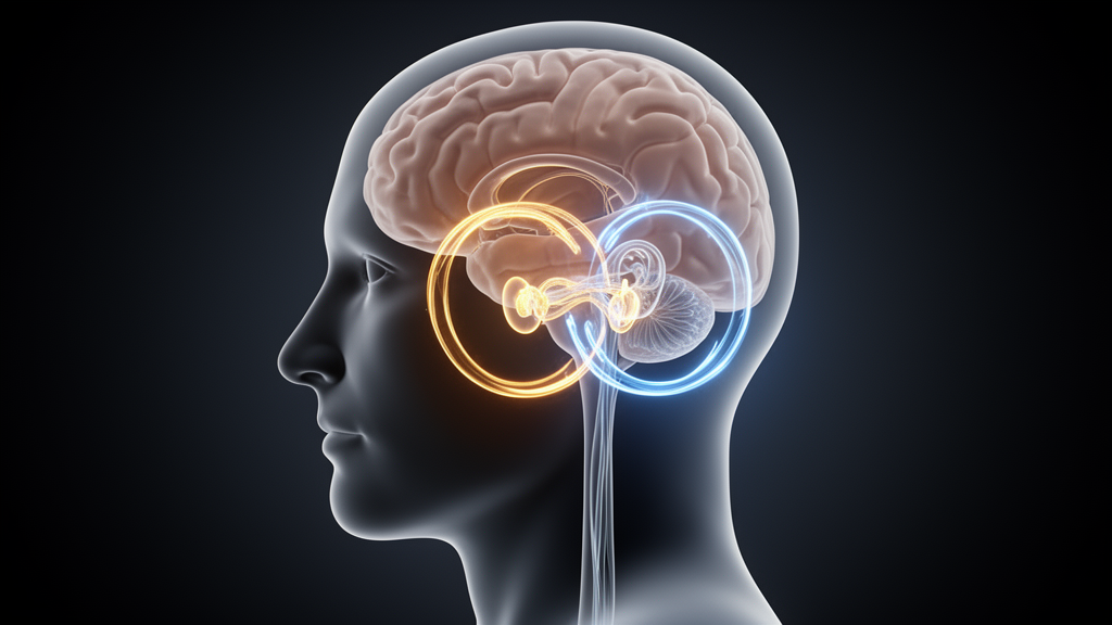
For most people, a spinning episode from an inner ear infection or displaced crystals fades within days as the brain rebalances its vestibular signals. For a substantial minority, the dizziness never fully leaves. A new review published in Frontiers in Integrative Neuroscience argues that this transition from acute vestibular event to chronic dizziness is driven not by the severity of the peripheral injury alone, but by two intertwined processes the authors call neural sensitization and psychological sensitization—a mutually reinforcing loop that locks symptoms in place.
The review, led by Moyan Wang of Changchun University of Chinese Medicine and colleagues at the Jilin Academy of Chinese Medicine Sciences, dissects the biology of vestibular compensation, the brain’s first-line recovery strategy. After unilateral vestibular injury, neurons in the ipsilateral vestibular nucleus lose their resting drive—a state of deafferentation that produces spontaneous nystagmus and postural deviation. Within hours, however, the intrinsic membrane properties of certain medial vestibular nucleus cells begin to change. B-type neurons, identifiable by their narrow action potentials and biphasic afterhyperpolarization, recover spontaneous firing as early as four hours after denervation through a compensatory increase in excitability, driven partly by bidirectional regulation of large-conductance calcium-activated potassium channels by CAMKII and PKC.
Static compensation—the rebalancing of resting discharge between the bilateral vestibular nuclei when the head is still—typically completes quickly. Dynamic compensation, the recovery of the vestibulo-ocular and vestibulospinal reflexes during head movement, is slower and often incomplete. During this later phase, long-term potentiation of synapses and commissural sprouting restore the sensitivity of deafferented neurons to head velocity and acceleration. Imaging studies using voxel-based morphometry and resting-state functional MRI reveal corresponding gray matter changes in the somatosensory cortex, superior temporal gyrus, insula, inferior parietal lobule, and posterior hippocampus, while animal work shows that brevican, a component of the perineuronal net in the superior vestibular nucleus, undergoes expression changes indicating extracellular matrix remodeling around neurons.
Yet this recovery carries a hidden cost: increased visual dependence. Sensory reweighting is the central nervous system’s dynamic adjustment of the relative contributions of vestibular, visual, and proprioceptive inputs to postural control. After a vestibular lesion, the weight of visual input rises while vestibular weight falls. Patients with bilateral vestibular dysfunction show markedly increased postural sway during visual or proprioceptive deprivation, yet perform nearly normally when both channels are intact. This dependence renders patients vulnerable to visual motion in complex environments—supermarket aisles, moving crowds, vehicular travel—producing visual vertigo in susceptible individuals.
Neural sensitization, the review’s first key mechanism, describes an abnormal increase in the processing gain of the vestibular system and its associated networks, causing normal stimuli to be perceived as abnormal vertigo. Synaptic remodeling provides the structural basis. Degradation of perineuronal nets may release constrained synaptic plasticity, promoting compensation while potentially permitting aberrant synaptic connections. Brain-derived neurotrophic factor, signaling through its TrkB receptor, drives remodeling of excitatory markers in the brainstem within the early window after vestibular neurectomy, balancing GABAergic neurons and GABA-A receptor expression between lesioned and intact sides—yet BDNF can also increase nervous system sensitivity by activating microglia. Glutamatergic synaptic strength in the ipsilateral medial vestibular nucleus, reflected by a decreased AMPA/NMDA ratio, reverses within twenty-four hours after labyrinthectomy, though presynaptic glutamate release probability remains incompletely compensated.
Neuroimaging in patients with persistent postural-perceptual dizziness and vestibular migraine substantiates these mechanisms. PPPD patients show significantly reduced gray matter volume in the inferior parietal lobule and hippocampus, negatively correlated with disease course—suggesting progressive structural depletion of key multisensory integration regions. Surface-based morphometry reveals reduced local gyrification in the bilateral posterior insular cortex, supramarginal gyrus, and posterior superior temporal gyrus, critical hubs for vestibular-somatosensory-visual convergence. During simulated vertical self-motion, activation of the parieto-insular vestibular cortex is markedly weaker in PPPD patients, while primary visual cortex activation intensity correlates positively with Dizziness Handicap Inventory scores—compensatory overactivation of the visual cortex. In vestibular migraine, regional homogeneity in the left middle temporal gyrus and middle occipital gyrus declines with increasing attack frequency.
Psychological sensitization, the second mechanism, operates through the fear-avoidance model, originally developed to explain chronic pain persistence. After a vertigo attack, patients develop catastrophic thinking about provoking movements—rapid head turns, turning around—believing these will inevitably cause a fall. Avoidance follows, and long-term avoidance alters proprioceptive input patterns from the neck, trunk, and lower limbs, making postural control more difficult and confirming the patient’s concern about instability. Neurobiologically, vestibular dysfunction activates the stress axis: upregulation of arginine vasopressin and corticotropin-releasing factor-immunoreactive cells in the paraventricular nucleus, and increased dopamine-β-hydroxylase in the locus coeruleus. The amygdala, detecting threat signals, sends excitatory projections to the locus coeruleus, which releases norepinephrine throughout the brain—including the vestibular nuclei—enhancing the spontaneous firing rate of medial vestibular nucleus neurons and their sensitivity to peripheral input. In chronic anxiety states, persistent amygdala overactivation keeps this system at high output, chronically elevating baseline vestibular excitability and reducing sensory thresholds.
Individual differences shape who descends this path. Neuroticism, assessed in a clinical observational study of 247 patients with chronic dizziness using the Eysenck Personality Questionnaire, was the only independent variable significantly predicting Dizziness Handicap Inventory scores, irrespective of diagnostic category and symptom duration. Neuroticism is associated with increased task-dependent functional connectivity between the amygdala and hippocampus and dorsolateral prefrontal cortex, and with altered coupling between the basolateral amygdala and anterior insular cortex—suggesting high-neuroticism individuals are more prone to hypervigilance after acute stress. Adverse childhood experiences produce long-term alterations in HPA axis function, while genetic variants including the BDNF Val66Met polymorphism, COMT Val158Met, and 5-HTTLPR short allele each modulate stress responsiveness, fear extinction learning, and anxiety susceptibility.
Crucially, the two sensitization processes do not operate independently. Neural sensitization lowers the brain’s processing threshold for vestibular, visual, and somatosensory information, increasing the frequency and salience of bodily signals—the subtle sway of normal posture and natural variations in visual scenes may be perceived as abnormal. Excessive bodily signals trigger hypervigilance and catastrophic appraisal. Psychological sensitization, in turn, enhances sensory processing gain through descending facilitatory pathways, modulated by limbic and autonomic systems. Patients who over-rely on visual input for spatial orientation experience longer-term vestibular symptoms; anxious patients show reduced proprioceptive thresholds and heightened sensitivity to body posture perception. Attentional orienting to proprioceptive stimulation alters processing in the primary sensorimotor cortex, decreasing beta wave power and increasing motor evoked potential amplitude.
The review proposes comprehensive interventions targeting sensory reweighting, fear-avoidance behavior, and psychiatric comorbidity to interrupt this process. Vestibular rehabilitation training, through repeated exposure to motion and visual stimuli that provoke dizziness, gradually reduces neural input sensitivity while breaking avoidance behaviors. Selective serotonin reuptake inhibitors and serotonin-norepinephrine reuptake inhibitors activate descending inhibitory pathways, reducing neural sensitivity. Transcranial alternating current stimulation of the dorsolateral prefrontal cortex effectively reduces vestibular responses and cortical excitability, while transcranial direct current stimulation targeting the parieto-insular vestibular cortex alleviates vestibular-related symptoms such as motion sickness. Antiepileptic drugs including pregabalin and topiramate inhibit calcium influx, reducing neural excitability. Cognitive behavioral therapy may benefit patients with pronounced fear-avoidance behavior and high Dizziness Handicap Inventory scores. The dual sensitization model remains a theoretical simplification—some individuals with severe health anxiety develop chronic dizziness-like symptoms without clear peripheral vestibular lesion, and most cited neuroimaging studies are cross-sectional with small samples—but it reframes chronic dizziness as a disorder of central processing gain rather than peripheral damage alone, offering a mechanistic bridge between the inner ear and the anxious brain.
Subject of Research: Mechanisms of the transition from acute vestibular events to chronic dizziness through neural and psychological sensitization
Article Title: Chronic dizziness: neuro-sensitization and psycho-sensitization
Article References: Wang, M., Han, Q., Teng, X., Liu, D., Yang, Z., Li, H., Yu, Y., Zhao, W., Xiang, K., & Sun, L. (2026). Chronic dizziness: neuro-sensitization and psycho-sensitization. Frontiers in Integrative Neuroscience, 20, Article 1929481. https://doi.org/10.3389/fnint.2026.1929481
Image Credits: AI Generated
DOI: 10.3389/fnint.2026.1929481
Keywords: chronic dizziness, neural sensitization, psychological sensitization, PPPD, vestibular migraine, vestibular compensation, sensory reweighting, fear-avoidance model, visual dependence, BDNF, amygdala, vestibular rehabilitation
News Source: Ophelia Keating. (October 16, 2026). How Acute Vertigo Becomes Chronic Dizziness: A Dual Sensitization Loop. Scienmag.