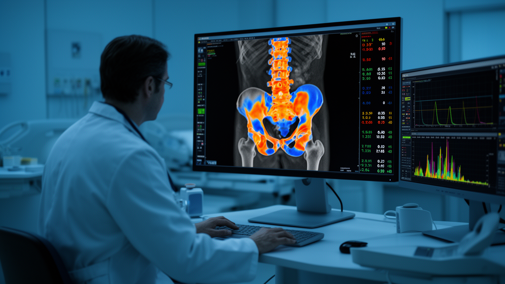
For decades, the nuclear medicine bone scan has been one of the most recognizable tests in imaging: a whole-body picture of glowing spots that reveals where the skeleton is actively remodeling. But its interpretation has always been fundamentally subjective, resting on the trained eye of a physician reading patterns of radiotracer uptake. A new review published in Frontiers in Nuclear Medicine argues that this is changing. Quantitative bone SPECT/CT, which converts the brightness of a lesion into standardized uptake values, or SUVs, is emerging as a way to measure bone metabolism objectively rather than describe it visually, and the technology is advancing quickly enough to challenge long-held assumptions about what a bone scan can do.
The review, conducted by Jovana Zivanovic of the University Clinical Centre of Serbia and Jules Zhang-Yin of Clinique Sud Luxembourg in Belgium, systematically searched the PubMed database for human studies published between January 2015 and March 2025 that applied quantitative or semi-quantitative analysis to bone SPECT/CT. Following PRISMA 2020 screening principles, the authors identified 150 records, whittled them down through title, abstract, and full-text assessment, and arrived at thirteen primary studies that formed the evidence base for a qualitative synthesis. Reviews, guidelines, and case reports were excluded from the core analysis but used for narrative context. The result is one of the most structured assessments to date of where quantitative bone SPECT/CT stands clinically and where it still falls short.
The technical foundation of the approach is straightforward in principle but demanding in practice. Quantification in SPECT/CT requires converting the counts detected by the gamma camera into absolute activity concentrations within tissue, a process that depends on a complete chain of corrections and calibrations. CT-based attenuation correction compensates for photons lost as they pass through the body, scatter correction removes contamination from deflected photons, and resolution recovery models partially undo the blurring introduced by the collimator. Cross-calibration between the dose calibrator and the camera, typically performed with phantoms, is essential, and recovery coefficients must be characterized for lesions of different sizes because partial-volume effects cause systematic underestimation in small lesions. Once these steps are in place, SUVmax, SUVmean, and SUVpeak can be calculated in a manner analogous to PET/CT, with SUVmax offering sensitivity to focal uptake at the cost of noise vulnerability, SUVmean providing stability but depending heavily on segmentation, and SUVpeak offering a compromise that may prove useful for multicentre follow-up studies.
The most mature clinical application lies in oncology, particularly in distinguishing malignant bone metastases from benign lesions. In one of the included studies, Du and colleagues found that malignant lesions showed significantly higher SUVmax than benign lesions and normal vertebrae, and that an SUVmax threshold of 18.07 identified malignancy with a reported prospective sensitivity of 82.35 percent, specificity of 93.75 percent, and accuracy of 89.80 percent. A separate study in lung adenocarcinoma by Lin and colleagues reported that a cutoff of 11.10 distinguished benign from malignant lesions with 87.70 percent sensitivity and 80.71 percent specificity, while a lower cutoff of 8.135 helped separate CT-negative metastases from normal vertebrae. Notably, the same study found that osteoblastic and mixed lesions had significantly greater SUVmax than osteolytic or CT-negative lesions, underscoring that lesion phenotype itself shapes the numbers.
Yet the review is emphatic that these thresholds cannot be treated as universal constants. Reported SUV cutoffs are influenced by tumour type, uptake time, acquisition and reconstruction parameters, calibration procedures, segmentation methodology, and recovery-coefficient handling, meaning that a value derived on one scanner with one protocol may be meaningless on another. The authors stress that quantitative SPECT/CT should currently be regarded as an adjunct to CT morphology, clinical information, and longitudinal imaging rather than a stand-alone diagnostic test. This caution is reinforced by the risk-of-bias appraisal performed for each of the thirteen studies, which found most to be of moderate quality, with several rated moderate to high risk in domains relating to confounding and external validation, reflecting the predominance of retrospective, single-centre designs with small and heterogeneous cohorts.
Beyond lesion characterization, quantitative bone SPECT/CT shows promise in tracking treatment response over time. Beck and colleagues demonstrated that longitudinal quantitative analysis of bone metabolism achieved almost perfect interobserver agreement, whereas visual analysis produced lower agreement, suggesting that numbers are more reproducible than eyeballs. Gherghe and colleagues showed that quantitative metrics could identify response patterns in metastatic breast cancer that were not always concordant with qualitative interpretation. Volumetric measures such as total lesion activity and skeletal tumour burden, analogous to metabolic tumour volume in FDG PET/CT, may provide global markers of disease extent, and one study by Dittmann and colleagues found that quantitative skeletal tumour burden and uptake parameters measured before radium-223 therapy were associated with patient outcomes, hinting at a prognostic role in bone-targeted radionuclide treatment.
The review also catalogues emerging applications in benign skeletal disease that are less widely appreciated. In mandibular condylar hyperplasia, a developmental deformity causing facial asymmetry, bone SPECT can detect whether the condyle is still actively growing by measuring uptake differences between the two sides, with a difference of roughly 10 percent or more on the affected side taken as evidence of active disease. In fibrous dysplasia, a rare disorder in which normal bone is replaced by immature fibro-osseous tissue, one study of 21 patients found that 18 showed moderate to high MDP metabolism, and a separate approach combining planar skeletal burden scores with lesion SUV values produced disease-corrected metrics that correlated with biological bone turnover markers. Quantitative SPECT/CT has also been applied to vertebral compression fractures, where Wang and colleagues identified an SUVmax cutoff of 9.925 for distinguishing fresh from old fractures, though a previous study reported a higher cutoff of 18.5, illustrating the transferability problem in miniature.
Normal reference values and patient-specific normalization represent another frontier. A study of 221 patients established baseline SUVmax values for normal cervical, thoracic, and lumbar vertebrae and found that males had significantly higher values than females across all three regions. Damian and colleagues investigated whether body weight, age, and CT-derived bone density could improve SUV normalization and found that standard body-weight SUV may overestimate uptake in heavier patients, while age and bone density significantly influenced vertebral uptake. Because many proposed thresholds for metastasis, fracture activity, or degenerative disease rest on SUVmax, wide interpatient variation in background bone uptake could render a single universal cutoff misleading, and future models may need to incorporate lean body mass, bone mineral density, renal function, hydration status, and tracer clearance.
On the hardware front, the review highlights the arrival of ring-configured cadmium-zinc-telluride, or CZT, detector systems that depart radically from the conventional rotating dual-head Anger camera. By arranging multiple stationary or semi-stationary detector modules in a 360-degree configuration around the patient, these systems capture a far greater proportion of emitted photons simultaneously, boosting sensitivity and count statistics. The gains translate into shorter acquisition times, lower administered activities, improved image quality, and better energy resolution that enhances scatter correction. An EANM position paper has argued that full-ring CZT bone imaging may fundamentally change the workflow of bone scintigraphy by making routine tomographic imaging feasible and reducing dependence on planar whole-body acquisitions. Dynamic SPECT acquisitions may even become possible, opening the door to kinetic modelling of tracer uptake rather than static snapshots.
Looking forward, the review identifies artificial intelligence, radiomics, and theranostics as research opportunities rather than established clinical applications. Radiomics could extract features beyond SUVmax and SUVmean to support automated lesion detection and response prediction, while deep-learning-enhanced reconstruction has already demonstrated the ability to maintain quantitative performance in ultra-fast acquisitions, potentially dissolving the traditional trade-off between scan time and image noise. In theranostics, quantitative SPECT/CT could support patient-specific dosimetry and treatment planning with widely available technetium-labelled tracers, though the authors caution that diphosphonate uptake reflects osteoblastic reaction and bone turnover rather than viable tumour burden alone. The overarching message is one of tempered optimism: quantitative bone SPECT/CT is technically feasible, increasingly reproducible, and clinically promising, but prospective multicentre studies with harmonized protocols, standardized calibration, and patient-relevant endpoints are needed before numbers can reliably replace the trained eye.
Subject of Research: Quantitative bone SPECT/CT imaging using standardized uptake values for skeletal disease assessment
Article Title: Quantification in bone SPECT/CT
Article References: Zivanovic, J., & Zhang-Yin, J. (2026). Quantification in bone SPECT/CT. Frontiers in Nuclear Medicine, 6, Article 1941906. https://doi.org/10.3389/fnume.2026.1941906
Image Credits: AI Generated
DOI: 10.3389/fnume.2026.1941906
Keywords: bone SPECT/CT, standardized uptake value, bone metastases, SUV quantification, CZT detectors, nuclear medicine, bone scintigraphy, vertebral compression fracture, fibrous dysplasia, radiomics, theranostics, SPECT reconstruction
News Source: Ophelia Keating. (October 16, 2026). Quantitative Bone SPECT/CT Moves From Visual Guesswork to Objective Numbers. Scienmag.