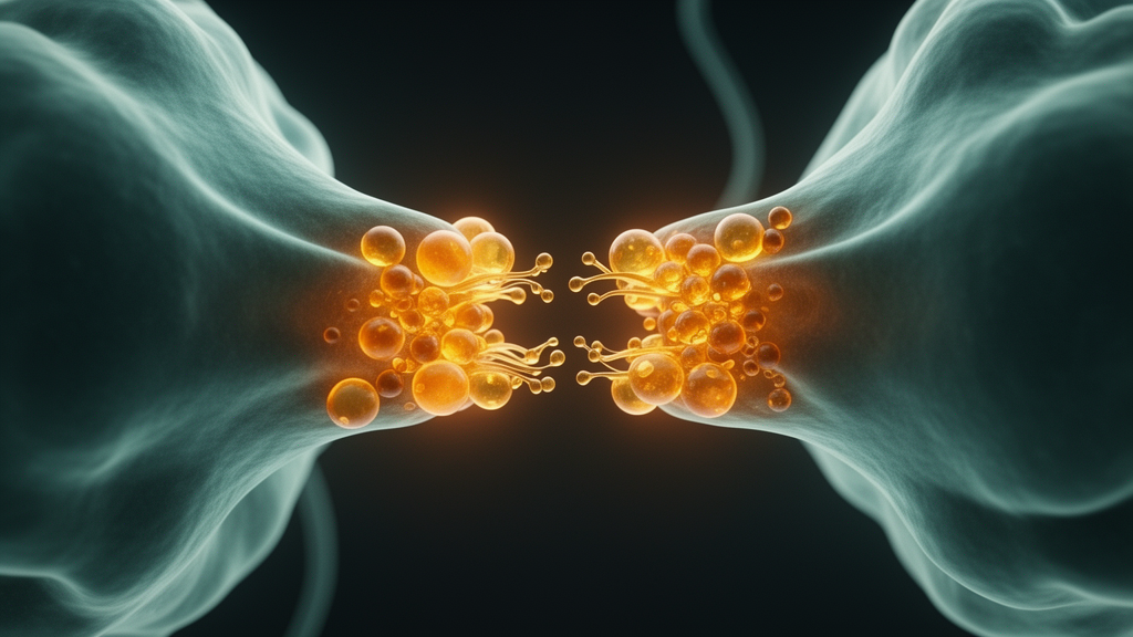
Synapses have long been described as the busiest construction sites in the brain, constantly dismantling and rebuilding their own membranes so that neurotransmitters can be packaged, released and recycled within milliseconds. A new review published in Frontiers in Cell Death argues that this same flexibility carries a hidden cost. Tetsumori Yamashima of Kanazawa University Graduate School of Medical Sciences proposes that when stresses such as ischemia, trauma or irradiation overwhelm presynaptic terminals, the lipid remodeling machinery that normally serves neurotransmission flips into a pathological mode, depositing lysophospholipids directly at the synapse and creating a form of regional lipotoxicity that standard lipidomics has largely missed.
The core of the argument rests on the peculiar biochemistry of synaptic membranes. Lipids account for up to 45 percent of the dry weight of the brain, and the presynaptic terminal is exceptionally rich in polyunsaturated fatty acids, particularly docosahexaenoic acid and arachidonic acid. Their carbon double bonds introduce curvature and conformational flexibility into the membrane, creating space between the hydrophobic core that allows receptors and ion channels to be interposed among phospholipids. This is precisely what makes rapid vesicle budding, fusion and neurotransmitter release possible. Yet the same unsaturated bonds are chemically vulnerable, prone to oxidative attack by reactive oxygen species that stresses generate in abundance.
Under physiological conditions, the enzyme phospholipase A2 cleaves membrane phosphatidylcholine in a controlled way, generating small amounts of lysophosphatidylcholine that help shape synaptic vesicles, while the enzyme lysophosphatidylcholine acyltransferase re-esterifies the molecule back into phosphatidylcholine through the Lands cycle. Excessive stimulation disrupts this equilibrium. Overactivated phospholipase A2 floods the presynaptic membrane with lysophosphatidylcholine, and the enzyme autotaxin converts much of it into lysophosphatidic acid. Both products distort membrane geometry: cylindrical phosphatidylcholine with two fatty acyl chains suits a flat bilayer, whereas the inverted-cone shape of single-chain lysophosphatidylcholine favors budding and, in excess, the formation of stacked lamellar structures.
The downstream consequences are severe. Lysophosphatidylcholine enhances membrane excitability at the synaptic cleft, while lysophosphatidic acid interferes with the cytoskeleton, triggering neurite retraction through increased phosphorylation of tau proteins. Free arachidonic acid released by phospholipase A2 becomes substrate for cyclooxygenases, yielding prostaglandins, thromboxanes and leukotrienes that amplify neuroinflammation. Meanwhile, lipid peroxidation of polyunsaturated membrane components and of mitochondrial cardiolipin produces the reactive aldehyde 4-hydroxy-2-nonenal, which carbonylates the lysosomal stabilizer heat-shock protein 70.1 and sets off a cascade of lysosomal rupture and neuronal death, a mechanism Yamashima’s group has detailed in previous work.
What has been missing, the review contends, is spatial resolution. Blood-based lipidomics can readily detect lysophosphatidylcholine and lysophosphatidic acid circulating in plasma, cerebrospinal fluid and other body fluids, and such measurements have documented elevations in Alzheimer’s disease, diabetes, ovarian cancer, renal failure and migraine. But these bulk assays homogenize tissue and lose regional information, and organic solvents used in extraction strip lysophosphatidylcholine out of brain samples. As a result, a pool of lysophospholipids concentrated within a few cubic millimeters of synaptic tissue, or within individual dendritic spines, remains effectively invisible to mass spectrometry even when it is locally toxic.
To close that gap, Yamashima revived and extended a fixation technique first introduced in 1972 by Mizuhira and Futaesaku. Tannic acid, a large galloylglucose molecule, interacts directly with the quaternary ammonium radical of the choline head group found in phosphatidylcholine and lysophosphatidylcholine, and the resulting aggregates are stabilized by postfixation with osmium tetroxide. Phospholipids lacking choline, such as phosphatidylethanolamine, phosphatidylinositol and phosphatidylserine, do not form these complexes. Under the electron microscope, intact phosphatidylcholine appears as distinct double membrane structures, while lysophosphatidylcholine reveals itself as extramembraneous, highly ordered multilamellar bodies with a repeating periodicity of roughly 45 angstroms.
The decisive experiment used Japanese macaque monkeys subjected to transient brain ischemia. In control neurons of the hippocampus, multilamellar structures were negligible at the synaptic area. In degenerating CA1 neurons, however, multilamellar bodies appeared clearly in the presynaptic regions from immediately after ischemia until day eight, in continuity with disintegrated presynaptic membranes and in the immediate vicinity of synaptic vesicles. The bodies appeared to enlarge by engulfing degenerating vesicle membranes, and their dependence on tannic acid staining marked them as choline-containing lysophosphatidylcholine, generated when overactivated phospholipase A2 cleaved the phosphatidylcholine of synaptic vesicle membranes. Even the ischemia-resistant granular neurons of the dentate gyrus deposited multilamellar phospholipids after the insult.
The review also connects these ultrastructural findings to a broader literature on lysophospholipid signaling. In mouse models of middle cerebral artery occlusion, mass spectrometry imaging showed phosphatidylcholine declining while lysophosphatidylcholine 16:0 rose and persisted for days in the affected hemisphere. Lysophosphatidic acid injected into the rat midbrain increased depression-like behavior in a dose-dependent manner, and blocking lysophosphatidic acid with a specific antibody reduced lesion volume and improved neurological outcomes after traumatic brain injury in mice. Patients with Alzheimer’s disease and mild cognitive impairment show lower plasma phosphatidylcholine and higher lysophosphatidylcholine, consistent with aberrant phospholipase A2 activity that amyloid beta 42 peptides are known to stimulate. Lysophosphatidylcholine also enhances the oligomer formation of amyloid beta 42, feeding directly into neurotoxic aggregation.
Importantly, the multilamellar bodies observed at synapses differ from those seen in lysosomal storage diseases such as Tay-Sachs and Niemann-Pick disease. In those disorders, lamellar accumulations sit within the neuronal cell soma, inside lysosomes packed with cholesterol and glycosphingolipids. The postischemic synaptic bodies, by contrast, appeared in dendritic spines at a distance from lysosome-like organelles, which concentrated in the perinuclear cytoplasm. Yamashima interprets them as breakdown products of synaptic vesicle membrane phospholipids rather than stored lipids, and notes that distorted lamella-like inclusions at synapses have been quietly visible in conventional electron micrographs of stressed and aged brains from rodents to humans, previously dismissed as fixation artifacts.
The practical implications reach toward the clinic. Because lysophosphatidic acid is water-soluble and present in vanishingly small amounts, it resists morphological detection, but its insoluble precursor lysophosphatidylcholine can now be visualized subcellularly. The author suggests that tiny one-to-three-millimeter cortical biopsies, obtained with informed consent during neurosurgical procedures for tumors, trauma, epilepsy or hydrocephalus, could be examined with tannic acid fixation to reveal lysophospholipid deposition in early human disease. If stress-induced lysophospholipid accumulation at synapses proves to be a common upstream event in cognitive and mental disorders, the transition from physiological membrane remodeling to pathological phospholipase A2 overactivation may become both a diagnostic marker and a therapeutic target for some of the most intractable diseases of the aging brain.
Subject of Research: Stress-induced synaptic accumulation of lysophospholipids and regional lipotoxicity in brain disease
Article Title: Synaptic deposition of lysophospholipids at stresses – risk of the regional lipotoxicity for brain diseases
Article References: Yamashima, T. (2026). Synaptic deposition of lysophospholipids at stresses – risk of the regional lipotoxicity for brain diseases. Frontiers in Cell Death, 5, Article 1858071. https://doi.org/10.3389/fceld.2026.1858071
Image Credits: AI Generated
DOI: 10.3389/fceld.2026.1858071
Keywords: lysophosphatidylcholine, lysophosphatidic acid, phospholipase A2, synapse, lipotoxicity, oxidative stress, electron microscopy, tannic acid, neurodegeneration, autotaxin, brain ischemia, lipidomics
News Source: Cassandra Pierce. (October 16, 2026). Stress Turns Brain Synapses Into Lipid Toxic Zones, New Review Warns. Scienmag.