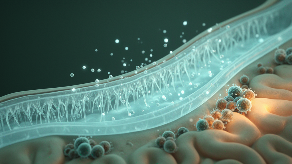
Deep inside the brain, along the walls of the fluid-filled ventricles, sits a thin lining of multiciliated cells that most people have never heard of: the ependyma. For decades, this delicate monolayer was treated as little more than biological wallpaper, a passive boundary between the cerebrospinal fluid and the brain tissue it bathes. A new study published in Nature Neuroscience argues that this underappreciated lining may be an active accomplice in one of the most stubborn features of multiple sclerosis, the diffuse degeneration that creeps inward from the ventricular surface and drives disability even in patients whose classic white matter lesions appear quiescent.
The research, led by Adam M. R. Groh and Jo Anne Stratton at the Montreal Neurological Institute-Hospital of McGill University, together with David A. Rudko and collaborators in Amsterdam and Montreal, set out to answer a question that has haunted the field since surface-in gradients of pathology were first described. Cerebrosp fluid-adjacent brain regions are especially vulnerable to diffuse degeneration in multiple sclerosis, and gradients of damage arising from cytotoxic factors carried in the cerebrospinal fluid have been proposed to predict disease relapses independently of white matter lesions. What remained unknown was whether those factors actually disrupt the ependymal cells that form the ventricular border, allowing them to penetrate into the brain parenchyma. The answer, according to the new work, is yes, and the mechanism involves a reactive, immune-responsive state in ependymal cells marked by the dysregulation of transporter and junctional proteins.
The technical achievement at the heart of the study is its multimodal design. The team used ultra-high-field 7-Tesla magnetic resonance imaging of ex vivo human brain tissue to reveal the presence of surface-in periventricular gradients in white matter, then guided histological sampling directly from the gradient-containing regions identified on the scans. This MRI-guided immunohistochemistry confirmed that gradient-containing regions show demyelination, increased microglial presence and axonal loss, and, critically, that they are lined by reactive ependymal cells. The approach solves a long-standing sampling problem in neuropathology: rather than choosing tissue blocks at random and hoping they capture the phenomenon of interest, the investigators let the imaging data dictate exactly where to look under the microscope.
Quantitatively, the histological findings were striking. Luxol fast blue staining, a standard measure of myelin integrity, showed significantly decreased mean pixel values in gradient-positive regions of multiple sclerosis tissue compared with both gradient-negative regions of the same patients and gradient-negative control tissue. The number of Iba1-positive microglia per square millimetre was increased in gradient-positive blocks, while axon counts per unit length were decreased. The team also documented increased numbers of wasteosomes, the corpora amylacea structures that accumulate in periventricular white matter hyperintensities, in gradient-positive regions. Together, these measurements establish that the periventricular gradient is not an imaging artifact but a reproducible histopathological reality with a defined molecular geography.
To understand what the ependymal cells themselves were doing, the researchers turned to single-nucleus multiomics, extracting nuclei from archived postmortem tissues and profiling both gene expression and chromatin accessibility. The single-nucleus RNA sequencing data confirmed the existence of a reactive transcriptomic state in human ependymal cells in multiple sclerosis, one strongly associated with dysregulation of cell-adhesion-associated genes. Gene ontology enrichment analyses revealed upregulation of terms related to cell junction assembly, and the team used SCENIC+, a computational framework for single-cell multiomic inference of enhancers and gene regulatory networks, to define the regulatory architecture underpinning the reactive state. Among the key transcription factors, STAT3 showed significantly increased expression and chromatin accessibility in a multiple sclerosis-associated ependymal cluster, while RFX3, a factor tied to ciliary biology, showed significantly decreased expression and accessibility.
The multiomic exploration went further, situating TNF and IFNγ as candidate drivers of the disease state. Using NicheNet, a method that models intercellular communication by linking ligands to target genes, the team predicted ligands enriched in multiple sclerosis cerebrospinal fluid that could drive the emergence of the reactive ependymal state. Electron microscopy of lesion-adjacent ependymal cells provided ultrastructural evidence of compromised cell adhesion: the investigators documented ependymal cells with disrupted cell-cell adhesion, lateral cellular processes protruding from detached cells, astrocyte processes invading the intercellular spaces formed by splitting ependymal cells, and, in the most severe cases, ependymal cells completely flattened or resorbed and replaced by a dense cobblestone-like network of astrocyte processes. These images capture the ventricular border in the act of failing.
The causal test came in vivo. The team acutely exposed murine ependymal cells to IFNγ by intracerebroventricular injection and found that the cytokine increased ependymal permeability, allowing a 500-kilodalton dextran tracer to infiltrate the brain parenchyma from the ventricular system. Three-dimensional reconstructions of whole mouse brains, generated through tissue clearing and light sheet microscopy as well as cryomicrotome imaging, showed that in vehicle-treated animals the dextran remained confined within the ventricles due to an intact ependymal monolayer, while after IFNγ exposure the tracer was observed infiltrating the periventricular region extensively. The effect was specific to the ependymal response: conditional knockout of ependymal Ifngr1, the gene encoding the IFNγ receptor subunit 1, prevented the increase in border permeability.
The in vivo experiments also revealed downstream inflammatory consequences. Intracerebroventricular IFNγ injection increased the number of periventricular CD45-positive immune cells compared with sham treatment, and conditional knockout of ependymal Ifngr1 reversed this effect, indicating that the ependymal response to the cytokine modulates regional inflammation rather than merely reflecting it. Notably, the number of CD45-positive cells in the choroid plexus showed no differences between experimental conditions, pointing to the ependymal lining itself, rather than the choroid plexus epithelium, as the relevant interface for this cytokine-driven recruitment. In vitro, the cytokine screen showed that IFNγ decreased normalized Foxj1 fluorescence intensity in ependymal cells, the master transcription factor of ciliated ependymal identity, while the combination of IFNγ and TNF reduced the percentage of Foxj1-positive cells, suggesting that combined exposure may impact ependymal cell survival.
The significance of the work extends beyond the ependyma. Periventricular magnetization transfer ratio gradients have been shown to occur early in multiple sclerosis and to improve after alemtuzumab treatment, and ependymal-in gradients of thalamic damage have been documented in progressive disease, where deep gray matter volume loss drives disability worsening. By identifying cerebrospinal fluid-driven ependymal dysfunction as a contributor to periventricular pathology, the study provides a cellular mechanism linking the fluid compartment to the tissue degeneration that standard lesion measures fail to capture. It also resonates with prior observations in neuromyelitis optica, where pathogenic cerebrospinal fluid barrier pathology has been described, and with the ependymal Dot-Dash sign, an early magnetic resonance imaging finding in multiple sclerosis that may now be interpretable as a visible trace of ependymal reactivity.
The data and code underpinning the study are openly available. The single-nucleus multiome datasets are deposited on GEO under accession GSE283715 and have been uploaded to the team’s open science platform SingloCell for further analysis, while all code used to analyze the ex vivo magnetic resonance images and the sequencing data is available on GitHub. The donors of the Douglas-Bell Canada Brain Bank made the human tissue work possible, and the murine experiments relied on Ifngr1-flanked, tamoxifen-inducible animals with tdTomato reporter expression to confirm ependymal-specific recombination. As the field moves toward mechanism-based criteria to improve therapeutic outcomes in progressive multiple sclerosis, the ventricular border has emerged from biological wallpaper to a pharmacologically addressable target, one whose integrity may determine how deeply the cerebrospinal fluid’s cytotoxic cargo penetrates the brain.
Subject of Research: Periventricular gradient pathology is linked to ependymal reactivity and cell adhesion disruption in multiple sclerosis
Article Title: Periventricular gradient pathology is linked to ependymal reactivity and cell adhesion disruption in multiple sclerosis
Article References: Periventricular gradient pathology is linked to ependymal reactivity and cell adhesion disruption in multiple sclerosis. (n.d.). https://doi.org/10.1038/s41593-026-02461-3
Image Credits: AI Generated
DOI: 10.1038/s41593-026-02461-3
Keywords: multiple sclerosis, ependymal cells, cerebrospinal fluid, neuroimmunology, single-nucleus multiomics, IFNγ, periventricular pathology, Nature Neuroscience, Periventricular, gradient, pathology, linked
News Source: Cassandra Pierce. (October 16, 2026). Cerebrospinal fluid signals breach the ventricular border in multiple sclerosis. Scienmag.